Chapter 9.11 Neurogenic pain – head and facial pain – neck pain With contributions by Jennifer Chu, Stuart Ferraris, Maureen Lovesey, 
|
Introduction NEUROGENIC PAIN | ||||||||||||||||||||||||||||||||||||||||||||||||||||||||||||||||||||||||||||||||||||||||||||||||||||||||||||||||||||||||||||||||||||||||||||||||||||||||||||||||||||||||||||||||||||||||||||||||||||||||||||||||||||||||||||||||||||||||||||||||||||||||||||||||||||||||||||||||||||||||||||||||||||||||||||||||||||||||||||||||||||||||||||||||||||||||||||||||||||||||||||||||||||||||||
|
The neuralgias | ||||||||||||||||||||||||||||||||||||||||||||||||||||||||||||||||||||||||||||||||||||||||||||||||||||||||||||||||||||||||||||||||||||||||||||||||||||||||||||||||||||||||||||||||||||||||||||||||||||||||||||||||||||||||||||||||||||||||||||||||||||||||||||||||||||||||||||||||||||||||||||||||||||||||||||||||||||||||||||||||||||||||||||||||||||||||||||||||||||||||||||||||||||||||||
|
Points used Parameters used | ||||||||||||||||||||||||||||||||||||||||||||||||||||||||||||||||||||||||||||||||||||||||||||||||||||||||||||||||||||||||||||||||||||||||||||||||||||||||||||||||||||||||||||||||||||||||||||||||||||||||||||||||||||||||||||||||||||||||||||||||||||||||||||||||||||||||||||||||||||||||||||||||||||||||||||||||||||||||||||||||||||||||||||||||||||||||||||||||||||||||||||||||||||||||||
|
Postherpetic neuralgia (PHN) | ||||||||||||||||||||||||||||||||||||||||||||||||||||||||||||||||||||||||||||||||||||||||||||||||||||||||||||||||||||||||||||||||||||||||||||||||||||||||||||||||||||||||||||||||||||||||||||||||||||||||||||||||||||||||||||||||||||||||||||||||||||||||||||||||||||||||||||||||||||||||||||||||||||||||||||||||||||||||||||||||||||||||||||||||||||||||||||||||||||||||||||||||||||||||||
|
Comparisons and combinations Points used When to use what – parameters for postherpetic neuralgia | ||||||||||||||||||||||||||||||||||||||||||||||||||||||||||||||||||||||||||||||||||||||||||||||||||||||||||||||||||||||||||||||||||||||||||||||||||||||||||||||||||||||||||||||||||||||||||||||||||||||||||||||||||||||||||||||||||||||||||||||||||||||||||||||||||||||||||||||||||||||||||||||||||||||||||||||||||||||||||||||||||||||||||||||||||||||||||||||||||||||||||||||||||||||||||
|
Acute herpetic neuralgia | ||||||||||||||||||||||||||||||||||||||||||||||||||||||||||||||||||||||||||||||||||||||||||||||||||||||||||||||||||||||||||||||||||||||||||||||||||||||||||||||||||||||||||||||||||||||||||||||||||||||||||||||||||||||||||||||||||||||||||||||||||||||||||||||||||||||||||||||||||||||||||||||||||||||||||||||||||||||||||||||||||||||||||||||||||||||||||||||||||||||||||||||||||||||||||
|
Comparisons and combinations Points used Parameters used | ||||||||||||||||||||||||||||||||||||||||||||||||||||||||||||||||||||||||||||||||||||||||||||||||||||||||||||||||||||||||||||||||||||||||||||||||||||||||||||||||||||||||||||||||||||||||||||||||||||||||||||||||||||||||||||||||||||||||||||||||||||||||||||||||||||||||||||||||||||||||||||||||||||||||||||||||||||||||||||||||||||||||||||||||||||||||||||||||||||||||||||||||||||||||||
|
Intercostal neuralgia | ||||||||||||||||||||||||||||||||||||||||||||||||||||||||||||||||||||||||||||||||||||||||||||||||||||||||||||||||||||||||||||||||||||||||||||||||||||||||||||||||||||||||||||||||||||||||||||||||||||||||||||||||||||||||||||||||||||||||||||||||||||||||||||||||||||||||||||||||||||||||||||||||||||||||||||||||||||||||||||||||||||||||||||||||||||||||||||||||||||||||||||||||||||||||||
|
Points used Parameters used | ||||||||||||||||||||||||||||||||||||||||||||||||||||||||||||||||||||||||||||||||||||||||||||||||||||||||||||||||||||||||||||||||||||||||||||||||||||||||||||||||||||||||||||||||||||||||||||||||||||||||||||||||||||||||||||||||||||||||||||||||||||||||||||||||||||||||||||||||||||||||||||||||||||||||||||||||||||||||||||||||||||||||||||||||||||||||||||||||||||||||||||||||||||||||||
|
Peripheral nerve injury and compression syndromes | ||||||||||||||||||||||||||||||||||||||||||||||||||||||||||||||||||||||||||||||||||||||||||||||||||||||||||||||||||||||||||||||||||||||||||||||||||||||||||||||||||||||||||||||||||||||||||||||||||||||||||||||||||||||||||||||||||||||||||||||||||||||||||||||||||||||||||||||||||||||||||||||||||||||||||||||||||||||||||||||||||||||||||||||||||||||||||||||||||||||||||||||||||||||||||
|
Comparisons and combinations Points used Parameters used | ||||||||||||||||||||||||||||||||||||||||||||||||||||||||||||||||||||||||||||||||||||||||||||||||||||||||||||||||||||||||||||||||||||||||||||||||||||||||||||||||||||||||||||||||||||||||||||||||||||||||||||||||||||||||||||||||||||||||||||||||||||||||||||||||||||||||||||||||||||||||||||||||||||||||||||||||||||||||||||||||||||||||||||||||||||||||||||||||||||||||||||||||||||||||||
|
The head and neck The shoulder and upper limb Thoracic outlet syndrome | ||||||||||||||||||||||||||||||||||||||||||||||||||||||||||||||||||||||||||||||||||||||||||||||||||||||||||||||||||||||||||||||||||||||||||||||||||||||||||||||||||||||||||||||||||||||||||||||||||||||||||||||||||||||||||||||||||||||||||||||||||||||||||||||||||||||||||||||||||||||||||||||||||||||||||||||||||||||||||||||||||||||||||||||||||||||||||||||||||||||||||||||||||||||||||
|
Points used Parameters used | ||||||||||||||||||||||||||||||||||||||||||||||||||||||||||||||||||||||||||||||||||||||||||||||||||||||||||||||||||||||||||||||||||||||||||||||||||||||||||||||||||||||||||||||||||||||||||||||||||||||||||||||||||||||||||||||||||||||||||||||||||||||||||||||||||||||||||||||||||||||||||||||||||||||||||||||||||||||||||||||||||||||||||||||||||||||||||||||||||||||||||||||||||||||||||
|
Compression syndrome of the lateral cutaneous forearm nerve | ||||||||||||||||||||||||||||||||||||||||||||||||||||||||||||||||||||||||||||||||||||||||||||||||||||||||||||||||||||||||||||||||||||||||||||||||||||||||||||||||||||||||||||||||||||||||||||||||||||||||||||||||||||||||||||||||||||||||||||||||||||||||||||||||||||||||||||||||||||||||||||||||||||||||||||||||||||||||||||||||||||||||||||||||||||||||||||||||||||||||||||||||||||||||||
|
Points used | ||||||||||||||||||||||||||||||||||||||||||||||||||||||||||||||||||||||||||||||||||||||||||||||||||||||||||||||||||||||||||||||||||||||||||||||||||||||||||||||||||||||||||||||||||||||||||||||||||||||||||||||||||||||||||||||||||||||||||||||||||||||||||||||||||||||||||||||||||||||||||||||||||||||||||||||||||||||||||||||||||||||||||||||||||||||||||||||||||||||||||||||||||||||||||
|
Ulnar nerve entrapment | ||||||||||||||||||||||||||||||||||||||||||||||||||||||||||||||||||||||||||||||||||||||||||||||||||||||||||||||||||||||||||||||||||||||||||||||||||||||||||||||||||||||||||||||||||||||||||||||||||||||||||||||||||||||||||||||||||||||||||||||||||||||||||||||||||||||||||||||||||||||||||||||||||||||||||||||||||||||||||||||||||||||||||||||||||||||||||||||||||||||||||||||||||||||||||
|
Points used | ||||||||||||||||||||||||||||||||||||||||||||||||||||||||||||||||||||||||||||||||||||||||||||||||||||||||||||||||||||||||||||||||||||||||||||||||||||||||||||||||||||||||||||||||||||||||||||||||||||||||||||||||||||||||||||||||||||||||||||||||||||||||||||||||||||||||||||||||||||||||||||||||||||||||||||||||||||||||||||||||||||||||||||||||||||||||||||||||||||||||||||||||||||||||||
|
Carpal tunnel syndrome (CTS) | ||||||||||||||||||||||||||||||||||||||||||||||||||||||||||||||||||||||||||||||||||||||||||||||||||||||||||||||||||||||||||||||||||||||||||||||||||||||||||||||||||||||||||||||||||||||||||||||||||||||||||||||||||||||||||||||||||||||||||||||||||||||||||||||||||||||||||||||||||||||||||||||||||||||||||||||||||||||||||||||||||||||||||||||||||||||||||||||||||||||||||||||||||||||||||
| Case study 9.11.1 (Ron Sharp) |   

| |||
| Comparisons and combinations | |||
| Points used | 
| |||
|
A caution | |||
|
Parameters used | |||
|
Plexus lesions Clunial nerve injury | ||||||||||||||||||||||||||||||||||||||||||||||||||||||||||||||||||||||||||||||||||||||||||||||||||||||||||||||||||||||||||||||||||||||||||||||||||||||||||||||||||||||||||||||||||||||||||||||||||||||||||||||||||||||||||||||||||||||||||||||||||||||||||||||||||||||||||||||||||||||||||||||||||||||||||||||||||||||||||||||||||||||||||||||||||||||||||||||||||||||||||||||||||||||||||
|
Points used Parameters used | ||||||||||||||||||||||||||||||||||||||||||||||||||||||||||||||||||||||||||||||||||||||||||||||||||||||||||||||||||||||||||||||||||||||||||||||||||||||||||||||||||||||||||||||||||||||||||||||||||||||||||||||||||||||||||||||||||||||||||||||||||||||||||||||||||||||||||||||||||||||||||||||||||||||||||||||||||||||||||||||||||||||||||||||||||||||||||||||||||||||||||||||||||||||||||
|
Piriformis syndrome | ||||||||||||||||||||||||||||||||||||||||||||||||||||||||||||||||||||||||||||||||||||||||||||||||||||||||||||||||||||||||||||||||||||||||||||||||||||||||||||||||||||||||||||||||||||||||||||||||||||||||||||||||||||||||||||||||||||||||||||||||||||||||||||||||||||||||||||||||||||||||||||||||||||||||||||||||||||||||||||||||||||||||||||||||||||||||||||||||||||||||||||||||||||||||||
|
Comparisons and combinations Points used Parameters used | ||||||||||||||||||||||||||||||||||||||||||||||||||||||||||||||||||||||||||||||||||||||||||||||||||||||||||||||||||||||||||||||||||||||||||||||||||||||||||||||||||||||||||||||||||||||||||||||||||||||||||||||||||||||||||||||||||||||||||||||||||||||||||||||||||||||||||||||||||||||||||||||||||||||||||||||||||||||||||||||||||||||||||||||||||||||||||||||||||||||||||||||||||||||||||
|
Meralgia paraesthetica Amputation pain | ||||||||||||||||||||||||||||||||||||||||||||||||||||||||||||||||||||||||||||||||||||||||||||||||||||||||||||||||||||||||||||||||||||||||||||||||||||||||||||||||||||||||||||||||||||||||||||||||||||||||||||||||||||||||||||||||||||||||||||||||||||||||||||||||||||||||||||||||||||||||||||||||||||||||||||||||||||||||||||||||||||||||||||||||||||||||||||||||||||||||||||||||||||||||||
|
Stump pain | ||||||||||||||||||||||||||||||||||||||||||||||||||||||||||||||||||||||||||||||||||||||||||||||||||||||||||||||||||||||||||||||||||||||||||||||||||||||||||||||||||||||||||||||||||||||||||||||||||||||||||||||||||||||||||||||||||||||||||||||||||||||||||||||||||||||||||||||||||||||||||||||||||||||||||||||||||||||||||||||||||||||||||||||||||||||||||||||||||||||||||||||||||||||||||
|
Comparisons and combinations Points used Parameters used | ||||||||||||||||||||||||||||||||||||||||||||||||||||||||||||||||||||||||||||||||||||||||||||||||||||||||||||||||||||||||||||||||||||||||||||||||||||||||||||||||||||||||||||||||||||||||||||||||||||||||||||||||||||||||||||||||||||||||||||||||||||||||||||||||||||||||||||||||||||||||||||||||||||||||||||||||||||||||||||||||||||||||||||||||||||||||||||||||||||||||||||||||||||||||||
|
Phantom pain | ||||||||||||||||||||||||||||||||||||||||||||||||||||||||||||||||||||||||||||||||||||||||||||||||||||||||||||||||||||||||||||||||||||||||||||||||||||||||||||||||||||||||||||||||||||||||||||||||||||||||||||||||||||||||||||||||||||||||||||||||||||||||||||||||||||||||||||||||||||||||||||||||||||||||||||||||||||||||||||||||||||||||||||||||||||||||||||||||||||||||||||||||||||||||||
|
Comparisons and combinations Points used When to use what – parameters for phantom pain | ||||||||||||||||||||||||||||||||||||||||||||||||||||||||||||||||||||||||||||||||||||||||||||||||||||||||||||||||||||||||||||||||||||||||||||||||||||||||||||||||||||||||||||||||||||||||||||||||||||||||||||||||||||||||||||||||||||||||||||||||||||||||||||||||||||||||||||||||||||||||||||||||||||||||||||||||||||||||||||||||||||||||||||||||||||||||||||||||||||||||||||||||||||||||||
|
Peripheral neuropathy | ||||||||||||||||||||||||||||||||||||||||||||||||||||||||||||||||||||||||||||||||||||||||||||||||||||||||||||||||||||||||||||||||||||||||||||||||||||||||||||||||||||||||||||||||||||||||||||||||||||||||||||||||||||||||||||||||||||||||||||||||||||||||||||||||||||||||||||||||||||||||||||||||||||||||||||||||||||||||||||||||||||||||||||||||||||||||||||||||||||||||||||||||||||||||||
|
Comparisons and combinations Points used When to use what – parameters for peripheral neuropathy | ||||||||||||||||||||||||||||||||||||||||||||||||||||||||||||||||||||||||||||||||||||||||||||||||||||||||||||||||||||||||||||||||||||||||||||||||||||||||||||||||||||||||||||||||||||||||||||||||||||||||||||||||||||||||||||||||||||||||||||||||||||||||||||||||||||||||||||||||||||||||||||||||||||||||||||||||||||||||||||||||||||||||||||||||||||||||||||||||||||||||||||||||||||||||||
|
Diabetic neuropathy | ||||||||||||||||||||||||||||||||||||||||||||||||||||||||||||||||||||||||||||||||||||||||||||||||||||||||||||||||||||||||||||||||||||||||||||||||||||||||||||||||||||||||||||||||||||||||||||||||||||||||||||||||||||||||||||||||||||||||||||||||||||||||||||||||||||||||||||||||||||||||||||||||||||||||||||||||||||||||||||||||||||||||||||||||||||||||||||||||||||||||||||||||||||||||||
|
Comparisons and combinations Points used Parameters used | ||||||||||||||||||||||||||||||||||||||||||||||||||||||||||||||||||||||||||||||||||||||||||||||||||||||||||||||||||||||||||||||||||||||||||||||||||||||||||||||||||||||||||||||||||||||||||||||||||||||||||||||||||||||||||||||||||||||||||||||||||||||||||||||||||||||||||||||||||||||||||||||||||||||||||||||||||||||||||||||||||||||||||||||||||||||||||||||||||||||||||||||||||||||||||
|
HIV-related peripheral neuropathy | ||||||||||||||||||||||||||||||||||||||||||||||||||||||||||||||||||||||||||||||||||||||||||||||||||||||||||||||||||||||||||||||||||||||||||||||||||||||||||||||||||||||||||||||||||||||||||||||||||||||||||||||||||||||||||||||||||||||||||||||||||||||||||||||||||||||||||||||||||||||||||||||||||||||||||||||||||||||||||||||||||||||||||||||||||||||||||||||||||||||||||||||||||||||||||
|
Points and parameters used | ||||||||||||||||||||||||||||||||||||||||||||||||||||||||||||||||||||||||||||||||||||||||||||||||||||||||||||||||||||||||||||||||||||||||||||||||||||||||||||||||||||||||||||||||||||||||||||||||||||||||||||||||||||||||||||||||||||||||||||||||||||||||||||||||||||||||||||||||||||||||||||||||||||||||||||||||||||||||||||||||||||||||||||||||||||||||||||||||||||||||||||||||||||||||||
|
Restless legs syndrome | ||||||||||||||||||||||||||||||||||||||||||||||||||||||||||||||||||||||||||||||||||||||||||||||||||||||||||||||||||||||||||||||||||||||||||||||||||||||||||||||||||||||||||||||||||||||||||||||||||||||||||||||||||||||||||||||||||||||||||||||||||||||||||||||||||||||||||||||||||||||||||||||||||||||||||||||||||||||||||||||||||||||||||||||||||||||||||||||||||||||||||||||||||||||||||
| A caution when treating peripheral neuropathy |
| |||||||||||||||||||||||||||||||||||||||||||||||||||||||||||||||||||||||||||||||||||||||||||||||||||||||||||||||||||||||||||||||||||||||||||||||||||||||||||||||||||||||||||||||||||||||||||||||||||||||||||||||||||||||||||||||||||||||||||||||||||||||||||||||||||||||||||||||||||||||||||||||||||||||||||||||||||||||||||||||||||||||||||||||||||||||||||||||||||||||||||||||||||||||||
|
Radiculopathy-related pain | ||||||||||||||||||||||||||||||||||||||||||||||||||||||||||||||||||||||||||||||||||||||||||||||||||||||||||||||||||||||||||||||||||||||||||||||||||||||||||||||||||||||||||||||||||||||||||||||||||||||||||||||||||||||||||||||||||||||||||||||||||||||||||||||||||||||||||||||||||||||||||||||||||||||||||||||||||||||||||||||||||||||||||||||||||||||||||||||||||||||||||||||||||||||||||
|
The low back and sciatica | ||||||||||||||||||||||||||||||||||||||||||||||||||||||||||||||||||||||||||||||||||||||||||||||||||||||||||||||||||||||||||||||||||||||||||||||||||||||||||||||||||||||||||||||||||||||||||||||||||||||||||||||||||||||||||||||||||||||||||||||||||||||||||||||||||||||||||||||||||||||||||||||||||||||||||||||||||||||||||||||||||||||||||||||||||||||||||||||||||||||||||||||||||||||||||
| Case study 9.11.2 (Juliette Lowe) |
| |||||||||||||||||||||||||||||||||||||||||||||||||||||||||||||||||||||||||||||||||||||||||||||||||||||||||||||||||||||||||||||||||||||||||||||||||||||||||||||||||||||||||||||||||||||||||||||||||||||||||||||||||||||||||||||||||||||||||||||||||||||||||||||||||||||||||||||||||||||||||||||||||||||||||||||||||||||||||||||||||||||||||||||||||||||||||||||||||||||||||||||||||||||||||
|
Comparisons and combinations Points used When to use what – parameters for radicular pain | ||||||||||||||||||||||||||||||||||||||||||||||||||||||||||||||||||||||||||||||||||||||||||||||||||||||||||||||||||||||||||||||||||||||||||||||||||||||||||||||||||||||||||||||||||||||||||||||||||||||||||||||||||||||||||||||||||||||||||||||||||||||||||||||||||||||||||||||||||||||||||||||||||||||||||||||||||||||||||||||||||||||||||||||||||||||||||||||||||||||||||||||||||||||||||
| Case study 9.11.3 (Jennifer Chu) |
| |||||||||||||||||||||||||||||||||||||||||||||||||||||||||||||||||||||||||||||||||||||||||||||||||||||||||||||||||||||||||||||||||||||||||||||||||||||||||||||||||||||||||||||||||||||||||||||||||||||||||||||||||||||||||||||||||||||||||||||||||||||||||||||||||||||||||||||||||||||||||||||||||||||||||||||||||||||||||||||||||||||||||||||||||||||||||||||||||||||||||||||||||||||||||
|
Complex regional pain disorder | ||||||||||||||||||||||||||||||||||||||||||||||||||||||||||||||||||||||||||||||||||||||||||||||||||||||||||||||||||||||||||||||||||||||||||||||||||||||||||||||||||||||||||||||||||||||||||||||||||||||||||||||||||||||||||||||||||||||||||||||||||||||||||||||||||||||||||||||||||||||||||||||||||||||||||||||||||||||||||||||||||||||||||||||||||||||||||||||||||||||||||||||||||||||||||
|
Points used When to use what – parameters for CRPD | ||||||||||||||||||||||||||||||||||||||||||||||||||||||||||||||||||||||||||||||||||||||||||||||||||||||||||||||||||||||||||||||||||||||||||||||||||||||||||||||||||||||||||||||||||||||||||||||||||||||||||||||||||||||||||||||||||||||||||||||||||||||||||||||||||||||||||||||||||||||||||||||||||||||||||||||||||||||||||||||||||||||||||||||||||||||||||||||||||||||||||||||||||||||||||
|
Central pain | ||||||||||||||||||||||||||||||||||||||||||||||||||||||||||||||||||||||||||||||||||||||||||||||||||||||||||||||||||||||||||||||||||||||||||||||||||||||||||||||||||||||||||||||||||||||||||||||||||||||||||||||||||||||||||||||||||||||||||||||||||||||||||||||||||||||||||||||||||||||||||||||||||||||||||||||||||||||||||||||||||||||||||||||||||||||||||||||||||||||||||||||||||||||||||
|
Pain following stroke Pain following spinal cord injury (SCI) Cauda equina syndrome, spinal stenosis and arachnoiditis | ||||||||||||||||||||||||||||||||||||||||||||||||||||||||||||||||||||||||||||||||||||||||||||||||||||||||||||||||||||||||||||||||||||||||||||||||||||||||||||||||||||||||||||||||||||||||||||||||||||||||||||||||||||||||||||||||||||||||||||||||||||||||||||||||||||||||||||||||||||||||||||||||||||||||||||||||||||||||||||||||||||||||||||||||||||||||||||||||||||||||||||||||||||||||||
|
Points used When to use what – parameters for central pain | ||||||||||||||||||||||||||||||||||||||||||||||||||||||||||||||||||||||||||||||||||||||||||||||||||||||||||||||||||||||||||||||||||||||||||||||||||||||||||||||||||||||||||||||||||||||||||||||||||||||||||||||||||||||||||||||||||||||||||||||||||||||||||||||||||||||||||||||||||||||||||||||||||||||||||||||||||||||||||||||||||||||||||||||||||||||||||||||||||||||||||||||||||||||||||
|
Scars and neurogenic pain | |||
|
HEAD AND FACIAL PAIN | ||||||||||||||||||||||||||||||||||||||||||||||||||||||||||||||||||||||||||||||||||||||||||||||||||||||||||||||||||||||||||||||||||||||||||||||||||||||||||||||||||||||||||||||||||||||||||||||||||||||||||||||||||||||||||||||||||||||||||||||||||||||||||||||||||||||||||||||||||||||||||||||||||||||||||||||||||||||||||||||||||||||||||||||||||||||||||||||||||||||||||||||||||||||||||
|
Headache | ||||||||||||||||||||||||||||||||||||||||||||||||||||||||||||||||||||||||||||||||||||||||||||||||||||||||||||||||||||||||||||||||||||||||||||||||||||||||||||||||||||||||||||||||||||||||||||||||||||||||||||||||||||||||||||||||||||||||||||||||||||||||||||||||||||||||||||||||||||||||||||||||||||||||||||||||||||||||||||||||||||||||||||||||||||||||||||||||||||||||||||||||||||||||||
|
Comparisons and combinations (general) Points used for headache – general considerations Parameters for headache – general considerations | ||||||||||||||||||||||||||||||||||||||||||||||||||||||||||||||||||||||||||||||||||||||||||||||||||||||||||||||||||||||||||||||||||||||||||||||||||||||||||||||||||||||||||||||||||||||||||||||||||||||||||||||||||||||||||||||||||||||||||||||||||||||||||||||||||||||||||||||||||||||||||||||||||||||||||||||||||||||||||||||||||||||||||||||||||||||||||||||||||||||||||||||||||||||||||
|
Tension-type headache | ||||||||||||||||||||||||||||||||||||||||||||||||||||||||||||||||||||||||||||||||||||||||||||||||||||||||||||||||||||||||||||||||||||||||||||||||||||||||||||||||||||||||||||||||||||||||||||||||||||||||||||||||||||||||||||||||||||||||||||||||||||||||||||||||||||||||||||||||||||||||||||||||||||||||||||||||||||||||||||||||||||||||||||||||||||||||||||||||||||||||||||||||||||||||||
|
Comparisons and combinations (general) Points used Parameters used | ||||||||||||||||||||||||||||||||||||||||||||||||||||||||||||||||||||||||||||||||||||||||||||||||||||||||||||||||||||||||||||||||||||||||||||||||||||||||||||||||||||||||||||||||||||||||||||||||||||||||||||||||||||||||||||||||||||||||||||||||||||||||||||||||||||||||||||||||||||||||||||||||||||||||||||||||||||||||||||||||||||||||||||||||||||||||||||||||||||||||||||||||||||||||||
|
Cluster headache | ||||||||||||||||||||||||||||||||||||||||||||||||||||||||||||||||||||||||||||||||||||||||||||||||||||||||||||||||||||||||||||||||||||||||||||||||||||||||||||||||||||||||||||||||||||||||||||||||||||||||||||||||||||||||||||||||||||||||||||||||||||||||||||||||||||||||||||||||||||||||||||||||||||||||||||||||||||||||||||||||||||||||||||||||||||||||||||||||||||||||||||||||||||||||||
|
Points used | ||||||||||||||||||||||||||||||||||||||||||||||||||||||||||||||||||||||||||||||||||||||||||||||||||||||||||||||||||||||||||||||||||||||||||||||||||||||||||||||||||||||||||||||||||||||||||||||||||||||||||||||||||||||||||||||||||||||||||||||||||||||||||||||||||||||||||||||||||||||||||||||||||||||||||||||||||||||||||||||||||||||||||||||||||||||||||||||||||||||||||||||||||||||||||
|
Other forms of headache | ||||||||||||||||||||||||||||||||||||||||||||||||||||||||||||||||||||||||||||||||||||||||||||||||||||||||||||||||||||||||||||||||||||||||||||||||||||||||||||||||||||||||||||||||||||||||||||||||||||||||||||||||||||||||||||||||||||||||||||||||||||||||||||||||||||||||||||||||||||||||||||||||||||||||||||||||||||||||||||||||||||||||||||||||||||||||||||||||||||||||||||||||||||||||||
|
Head and facial pains of vascular origin | ||||||||||||||||||||||||||||||||||||||||||||||||||||||||||||||||||||||||||||||||||||||||||||||||||||||||||||||||||||||||||||||||||||||||||||||||||||||||||||||||||||||||||||||||||||||||||||||||||||||||||||||||||||||||||||||||||||||||||||||||||||||||||||||||||||||||||||||||||||||||||||||||||||||||||||||||||||||||||||||||||||||||||||||||||||||||||||||||||||||||||||||||||||||||||
|
Migraine | ||||||||||||||||||||||||||||||||||||||||||||||||||||||||||||||||||||||||||||||||||||||||||||||||||||||||||||||||||||||||||||||||||||||||||||||||||||||||||||||||||||||||||||||||||||||||||||||||||||||||||||||||||||||||||||||||||||||||||||||||||||||||||||||||||||||||||||||||||||||||||||||||||||||||||||||||||||||||||||||||||||||||||||||||||||||||||||||||||||||||||||||||||||||||||
|
Comparisons and combinations Points used Parameters used | ||||||||||||||||||||||||||||||||||||||||||||||||||||||||||||||||||||||||||||||||||||||||||||||||||||||||||||||||||||||||||||||||||||||||||||||||||||||||||||||||||||||||||||||||||||||||||||||||||||||||||||||||||||||||||||||||||||||||||||||||||||||||||||||||||||||||||||||||||||||||||||||||||||||||||||||||||||||||||||||||||||||||||||||||||||||||||||||||||||||||||||||||||||||||||
|
Non-migrainous vascular headache | ||||||||||||||||||||||||||||||||||||||||||||||||||||||||||||||||||||||||||||||||||||||||||||||||||||||||||||||||||||||||||||||||||||||||||||||||||||||||||||||||||||||||||||||||||||||||||||||||||||||||||||||||||||||||||||||||||||||||||||||||||||||||||||||||||||||||||||||||||||||||||||||||||||||||||||||||||||||||||||||||||||||||||||||||||||||||||||||||||||||||||||||||||||||||||
|
Horton’s headache Charlin’s syndrome Sluder’s syndrome | ||||||||||||||||||||||||||||||||||||||||||||||||||||||||||||||||||||||||||||||||||||||||||||||||||||||||||||||||||||||||||||||||||||||||||||||||||||||||||||||||||||||||||||||||||||||||||||||||||||||||||||||||||||||||||||||||||||||||||||||||||||||||||||||||||||||||||||||||||||||||||||||||||||||||||||||||||||||||||||||||||||||||||||||||||||||||||||||||||||||||||||||||||||||||||
|
Comparisons and combinations for non-migrainous vascular headache Points used for non-migrainous vascular headache Parameters used for non-migrainous vascular headache | ||||||||||||||||||||||||||||||||||||||||||||||||||||||||||||||||||||||||||||||||||||||||||||||||||||||||||||||||||||||||||||||||||||||||||||||||||||||||||||||||||||||||||||||||||||||||||||||||||||||||||||||||||||||||||||||||||||||||||||||||||||||||||||||||||||||||||||||||||||||||||||||||||||||||||||||||||||||||||||||||||||||||||||||||||||||||||||||||||||||||||||||||||||||||||
|
Facial pain | ||||||||||||||||||||||||||||||||||||||||||||||||||||||||||||||||||||||||||||||||||||||||||||||||||||||||||||||||||||||||||||||||||||||||||||||||||||||||||||||||||||||||||||||||||||||||||||||||||||||||||||||||||||||||||||||||||||||||||||||||||||||||||||||||||||||||||||||||||||||||||||||||||||||||||||||||||||||||||||||||||||||||||||||||||||||||||||||||||||||||||||||||||||||||||
|
Comparisons and combinations – facial pain in general Points used for facial pain – facial pain in general Parameters used for facial pain – facial pain in general | ||||||||||||||||||||||||||||||||||||||||||||||||||||||||||||||||||||||||||||||||||||||||||||||||||||||||||||||||||||||||||||||||||||||||||||||||||||||||||||||||||||||||||||||||||||||||||||||||||||||||||||||||||||||||||||||||||||||||||||||||||||||||||||||||||||||||||||||||||||||||||||||||||||||||||||||||||||||||||||||||||||||||||||||||||||||||||||||||||||||||||||||||||||||||||
|
Trigeminal neuralgia, or tic douloureux | ||||||||||||||||||||||||||||||||||||||||||||||||||||||||||||||||||||||||||||||||||||||||||||||||||||||||||||||||||||||||||||||||||||||||||||||||||||||||||||||||||||||||||||||||||||||||||||||||||||||||||||||||||||||||||||||||||||||||||||||||||||||||||||||||||||||||||||||||||||||||||||||||||||||||||||||||||||||||||||||||||||||||||||||||||||||||||||||||||||||||||||||||||||||||||
|
Comparisons and combinations Points used Parameters used | ||||||||||||||||||||||||||||||||||||||||||||||||||||||||||||||||||||||||||||||||||||||||||||||||||||||||||||||||||||||||||||||||||||||||||||||||||||||||||||||||||||||||||||||||||||||||||||||||||||||||||||||||||||||||||||||||||||||||||||||||||||||||||||||||||||||||||||||||||||||||||||||||||||||||||||||||||||||||||||||||||||||||||||||||||||||||||||||||||||||||||||||||||||||||||
|
Atypical facial pain | ||||||||||||||||||||||||||||||||||||||||||||||||||||||||||||||||||||||||||||||||||||||||||||||||||||||||||||||||||||||||||||||||||||||||||||||||||||||||||||||||||||||||||||||||||||||||||||||||||||||||||||||||||||||||||||||||||||||||||||||||||||||||||||||||||||||||||||||||||||||||||||||||||||||||||||||||||||||||||||||||||||||||||||||||||||||||||||||||||||||||||||||||||||||||||
|
Comparisons and combinations Points used Parameters used | ||||||||||||||||||||||||||||||||||||||||||||||||||||||||||||||||||||||||||||||||||||||||||||||||||||||||||||||||||||||||||||||||||||||||||||||||||||||||||||||||||||||||||||||||||||||||||||||||||||||||||||||||||||||||||||||||||||||||||||||||||||||||||||||||||||||||||||||||||||||||||||||||||||||||||||||||||||||||||||||||||||||||||||||||||||||||||||||||||||||||||||||||||||||||||
|
Occipital neuralgia | ||||||||||||||||||||||||||||||||||||||||||||||||||||||||||||||||||||||||||||||||||||||||||||||||||||||||||||||||||||||||||||||||||||||||||||||||||||||||||||||||||||||||||||||||||||||||||||||||||||||||||||||||||||||||||||||||||||||||||||||||||||||||||||||||||||||||||||||||||||||||||||||||||||||||||||||||||||||||||||||||||||||||||||||||||||||||||||||||||||||||||||||||||||||||||
|
Points used Parameters used | ||||||||||||||||||||||||||||||||||||||||||||||||||||||||||||||||||||||||||||||||||||||||||||||||||||||||||||||||||||||||||||||||||||||||||||||||||||||||||||||||||||||||||||||||||||||||||||||||||||||||||||||||||||||||||||||||||||||||||||||||||||||||||||||||||||||||||||||||||||||||||||||||||||||||||||||||||||||||||||||||||||||||||||||||||||||||||||||||||||||||||||||||||||||||||
|
Arnold’s neuralgia Temporomandibular joint dysfunction | ||||||||||||||||||||||||||||||||||||||||||||||||||||||||||||||||||||||||||||||||||||||||||||||||||||||||||||||||||||||||||||||||||||||||||||||||||||||||||||||||||||||||||||||||||||||||||||||||||||||||||||||||||||||||||||||||||||||||||||||||||||||||||||||||||||||||||||||||||||||||||||||||||||||||||||||||||||||||||||||||||||||||||||||||||||||||||||||||||||||||||||||||||||||||||
|
Comparisons and combinations Points used | |||
| Caution | | |||||||||||||||||||||||||||||||||||||||||||||||||||||||||||||||||||||||||||||||||||||||||||||||||||||||||||||||||||||||||||||||||||||||||||||||||||||||||||||||||||||||||||||||||||||||||||||||||||||||||||||||||||||||||||||||||||||||||||||||||||||||||||||||||||||||||||||||||||||||||||||||||||||||||||||||||||||||||||||||||||||||||||||||||||||||||||||||||||||||||||||||||||||||||
|
Parameters used | ||||||||||||||||||||||||||||||||||||||||||||||||||||||||||||||||||||||||||||||||||||||||||||||||||||||||||||||||||||||||||||||||||||||||||||||||||||||||||||||||||||||||||||||||||||||||||||||||||||||||||||||||||||||||||||||||||||||||||||||||||||||||||||||||||||||||||||||||||||||||||||||||||||||||||||||||||||||||||||||||||||||||||||||||||||||||||||||||||||||||||||||||||||||||||
|
Dental pain | ||||||||||||||||||||||||||||||||||||||||||||||||||||||||||||||||||||||||||||||||||||||||||||||||||||||||||||||||||||||||||||||||||||||||||||||||||||||||||||||||||||||||||||||||||||||||||||||||||||||||||||||||||||||||||||||||||||||||||||||||||||||||||||||||||||||||||||||||||||||||||||||||||||||||||||||||||||||||||||||||||||||||||||||||||||||||||||||||||||||||||||||||||||||||||
|
Box 9.11.1 Some conclusions from experimental studies | ||||||||||||||||||||||||||||||||||||||||||||||||||||||||||||||||||||||||||||||||||||||||||||||||||||||||||||||||||||||||||||||||||||||||||||||||||||||||||||||||||||||||||||||||||||||||||||||||||||||||||||||||||||||||||||||||||||||||||||||||||||||||||||||||||||||||||||||||||||||||||||||||||||||||||||||||||||||||||||||||||||||||||||||||||||||||||||||||||||||||||||||||||||||||||
| Case study 9.11.4 (Stuart Ferraris) |   
| |||||||||||||||||||||||||||||||||||||||||||||||||||||||||||||||||||||||||||||||||||||||||||||||||||||||||||||||||||||||||||||||||||||||||||||||||||||||||||||||||||||||||||||||||||||||||||||||||||||||||||||||||||||||||||||||||||||||||||||||||||||||||||||||||||||||||||||||||||||||||||||||||||||||||||||||||||||||||||||||||||||||||||||||||||||||||||||||||||||||||||||||||||||||||
|
Comparisons and combinations Points used Parameters used | ||||||||||||||||||||||||||||||||||||||||||||||||||||||||||||||||||||||||||||||||||||||||||||||||||||||||||||||||||||||||||||||||||||||||||||||||||||||||||||||||||||||||||||||||||||||||||||||||||||||||||||||||||||||||||||||||||||||||||||||||||||||||||||||||||||||||||||||||||||||||||||||||||||||||||||||||||||||||||||||||||||||||||||||||||||||||||||||||||||||||||||||||||||||||||
|
NECK PAIN | ||||||||||||||||||||||||||||||||||||||||||||||||||||||||||||||||||||||||||||||||||||||||||||||||||||||||||||||||||||||||||||||||||||||||||||||||||||||||||||||||||||||||||||||||||||||||||||||||||||||||||||||||||||||||||||||||||||||||||||||||||||||||||||||||||||||||||||||||||||||||||||||||||||||||||||||||||||||||||||||||||||||||||||||||||||||||||||||||||||||||||||||||||||||||||
| Case study 9.11.5 (Maureen Lovesey) |
| |||||||||||||||||||||||||||||||||||||||||||||||||||||||||||||||||||||||||||||||||||||||||||||||||||||||||||||||||||||||||||||||||||||||||||||||||||||||||||||||||||||||||||||||||||||||||||||||||||||||||||||||||||||||||||||||||||||||||||||||||||||||||||||||||||||||||||||||||||||||||||||||||||||||||||||||||||||||||||||||||||||||||||||||||||||||||||||||||||||||||||||||||||||||||
|
Comparisons and combinations Points used Parameters used | ||||||||||||||||||||||||||||||||||||||||||||||||||||||||||||||||||||||||||||||||||||||||||||||||||||||||||||||||||||||||||||||||||||||||||||||||||||||||||||||||||||||||||||||||||||||||||||||||||||||||||||||||||||||||||||||||||||||||||||||||||||||||||||||||||||||||||||||||||||||||||||||||||||||||||||||||||||||||||||||||||||||||||||||||||||||||||||||||||||||||||||||||||||||||||
|
Summary Clinical studies database: Summary chart 9.11 | |||
| Other icons used in this chapter: |
    
| |
In this subchapter, treatments for neurogenic pain, head and facial pain, and neck pain are described. The focus is on electrical stimulation, but some information is included on laser acupuncture (LA) and low-intensity laser therapy (LILT), which have been used for many of the conditions covered. Neurogenic pain may be due to nerve injury, neuritis (nerve inflammation) or degenerative (non-inflammatory) neuropathy, peripheral or central in origin. Some neurogenic pain may accompany or follow infection.1 It can take the form of neuralgia (paroxysmal or fulminating pain along the course of one or more nerves), or be less severe but prolonged, sometimes with a correspondingly increased or decreased excitability to electrical methods of stimulation.2 Neurogenic paraesthesiae (distorted sensations) may include burning, ‘pins and needles’ or numbness. Neurogenic pain may be associated with reduced variability of electrical activity in the thalamus (ventral caudal nucleus), but also increased activity (frequency) in other thalamic regions (see SubCh. 5.1). Interestingly, referred sensations on electrical stimulation of ear points are not uncommon with neurogenic pain.3 Acupuncture has been used for neuralgia in the West since at least the 1850s.4
Microwave acupuncture has been used for neuralgia, both in humans9 and in animals (see Ch. 10, ‘used for neuralgia’). Ultrasound acupuncture has been used for neuralgic pain.10 Anton Jayasuriya has found that auricular LA can be helpful for neuralgia.11 Voll, in his EAV system, proposed that 3.9 Hz acupoint stimulation should generally be used for neuralgia, although it is unclear why he adopted this particular frequency.12 In a review of TENS for pain, Thomas Lundeberg commented that neuralgia tends to respond well to CTENS.13 Herpes zoster and its treatments are described elsewhere (see SubCh. 9.4). Following the acute stage, postherpetic neuralgia can develop if the virus has not been adequately contained.14 This is the most common and dreaded complication of herpes zoster.15 Although PHN resolves spontaneously within 3 months in around 50% of cases, it may persist in 14%–22%.16,17 Like herpes zoster itself, PHN tends to be more of a problem in older people (it is uncommon in those under 60 years18), and once it has become chronic can be very difficult to treat whatever method is used. Conventional treatment includes sympathetic blockade,19 antidepressant medication such as amitriptyline and the anticonvulsant carbamazepine,20 although other drugs such as the NMDA blockers ketamine and simple magnesium have also been investigated.21,22 As so often, the quality of many studies and reviews of conventional treatments for PHN has been criticised.23 The conclusion of one systematic review of RCTs for PHN was that tricyclic antidepressants appear to be the only agents of proven benefit once PHN is established.24 Preventive use of tricyclics at the onset of the acute phase has also been attempted,25 and virustatics such as acyclovir may reduce time to pain relief26 if not altogether prevent subsequent PHN.27 Overmedication is a risk in the elderly.28 Topical agents such as capsaicin and lidocaine may be helpful according to one informal review.29 PHN most commonly affects the ophthalmic division of the trigeminal nerve (see SubCh. 9.4) and midthoracic dermatomes, with least probability of pain relief for PHN of the isolated ophthalmic nerve and of the brachial plexus, and greatest when it involves the jaw, neck and trunk.30 The pain experienced may be both steady and paroxysmal. Sensory loss, allodynia and dysaesthesia (disturbances of sensation) are also common, whereas hyperalgesia (pain of abnormal severity following noxious stimulation31) is less so. There may be a better chance of recovery if allodynia or sensory deficit is absent.32
Early treatment of acute herpes zoster is important in reducing the likelihood of subsequent PHN (see SubCh. 9.4, ‘24–72 hours’). Provided treatment is started early following the initial acute phase, EA and TENS may also be of benefit once PHN has started. Given that intravenous magnesium can temporarily block the NMDA receptor and alleviate PHN, it would be interesting to see whether oral magnesium supplements could alleviate PHN and other types of neurogenic pain,81 particularly in conjunction with acupuncture, which may itself increase blood magnesium level.82 As mentioned, although MA is probably not very effective for PHN, TENS, and particularly CTENS, has often been considered helpful.83 However, in one small RCT, EA (5/60 Hz DD) for 6 weeks gave better results than CTENS.84 Pain in the EA group worsened again after treatment was terminated, so some commentators have surmised that the increased attention received by these patients was a major factor in their improvement (also that the different stimulation parameters used contributed in some way). In another study in which both EA (‘muscle stimulation’ with needles) and TENS were used, EA was often helpful in patients with dermatomal lack of sensation (so CTENS was not appropriate). Again, though, continued treatment was necessary for extended pain relief.85 EA has been combined with cupping for PHN.86 The results of this study are fascinating if difficult to interpret: 100% of the 25 Chinese patients treated were reported as cured, but only 6.2% of German patients, and 20% of the latter did not respond at all! LA was considered superior to MA in one small comparative study.87 Some patients who did not respond to LA alone did better with a combination of both methods. Similarly, the combination of HeNe LA with simultaneous magnetic field treatment has been claimed to give better results than LA alone.88 Keith Glennie-Smith has suggested that segmental acupuncture may not be effective because myelinated afferents have been preferentially destroyed by the herpes zoster virus, and that points rostral or contralateral to the affected dermatome or auricular points should be treated instead (although, curiously, he also advocates ‘surrounding the dragon’ locally as an effective method).89 For the same reason, deep-muscle stimulation may be more effective than superficial dermatomal treatment.90 Paravertebral (or huatuojiaji) points at the level of the lesion are recommended by George Ulett among others.91,92 Points that appear several times in the studies in the database ( ST-36 has been described as the most frequently selected point for PHN in Chinese studies,93 although I understand that in one ‘mixed’ TCM and Western approach taught in the UK distal points such as GB-34 and LIV-3 are recommended, with ‘local’ points above and below the affected dermatome. Electrode placements in the TENS studies of PHN include trigger points, selected dermatomes, low electrical resistance acupuncture points, over the painful area and proximal to the pain.94 John Thompson and Jacqueline Filshie suggest electrodes close to but not directly over the affected area in the same or adjacent dermatomes,95 possibly straddling the affected dermatome.96 Contralateral TENS may be an alternative.97 In practice, Glennie-Smith uses LILT locally as well as over intervertebral points, directed at the posterior root ganglion and dorsal horn at the affected spinal level (as well as in dermatomes immediately above and below it).98 Another approach is to irradiate the stellate ganglion.99 When to use what – parameters for postherpetic neuralgia PHN did not respond well to LF EA in one retrospective study,100 although LF EA did give good results in Chinese (but not German) patients in another uncontrolled trial.101 However, 5/60 Hz DD EA did give better results than CTENS at least in the short term (see above for a brief discussion of this comparison). In the PHN TENS studies, parameters varied widely: stimulation frequency from 10 to 180 Hz, intensities from ‘low’ to comfortable to the maximum tolerated, with duration from 20 minutes daily to ‘prolonged’. Even with this limited data, there does appear to be some evidence that high-intensity 100 Hz stimulation long term may be of benefit.102 As always, though, more research is necessary to confirm this.103 Margareta Eriksson and colleagues, on the basis of a small uncontrolled study, considered that HF or burst TENS gave better results for PHN than ALTENS104 ( Keith Glennie-Smith has used red LILT (660 nm) for local superficial irradiation (contact mode, 3 W/cm2, 8–10 seconds/pt), with the same dose (24–30 J/cm2) of infrared LILT (820–840 nm, 50 mW, 60–70 Hz) for deeper penetration at intervertebral points107 (904 nm may also be an appropriate wavelength108). Higher output power (150 mW rather than 60 mW) may be more effective still.109 Glennie-Smith suggests twice-weekly treatments initially, whereas Boris Sommer, writing more recently, recommends a more intensive approach, with five to six daily sessions initially, slowly reducing treatment frequency to once weekly.110 As emphasised above, it is important to treat herpetic pain early if it is not to become chronic. MA111 and EA112,113 have been used for acute herpetic pain, with a reduced incidence of PHN with the latter in two studies.114,115
In one report on acupuncture for acute herpes zoster pain, EA gave better results than MA ‘in some cases’.123 As with acute herpes zoster in general, the local lesions are often the focus of treatment, using the ‘surround the dragon’ technique, for example (see Figs. 9.6.1, 10.1c). Posterior nerve roots (or huatuojiaji points) in the same segment can be used, and in one report anterior segmental points were used as well.124 A similar approach was used in the ‘PENS’ (EA) study by Hesham Ahmed and colleagues.125 However, rather than treating the affected segment itself, stimulation was limited to the dermatomes above and below it. Of the traditional acupoints, GB-43, LIV-3 and LI-4 have been used in more than one study. LF and HF and 10 Hz stimulation have been used for acute herpes zoster pain. In one interesting report, HF EA was used first (for analgesia), followed by LF EA (for its neurotrophic effect).126 With LA/LILT, HeNe is common, although polarised non-laser irradiation has also been used.127 (For craniofacial neuralgias other than PHN, and for occipital and cervicobrachial neuralgia, see below, ‘Facial pain’, and ‘Occipital neuralgia’ and ‘Neck pain’, respectively).
Magnets positioned at acupoints have been used for intercostal neuralgia.133,134 George Ulett recommends using paravertebral points at the level of the lesion involved, as with PHN.135 Apart from the huatuojiaji or back shu points (BL-17, BL-18), others such as GB-34, LIV-3 and LIV-14 have also been suggested,136 or LIV-3 and LIV-14 with other distal points such as SJ-6 and SP-9 or ST-40 when the condition is considered as due to stagnation of Liver qi.137 P-6 has been used for acute intercostal neuralgia.138 In one EA device manual, HF or DD is recommended for intercostal neuralgia, at a moderate to strong intensity.139 Another just favours DD.140 Peripheral nerve injury and compression syndromes Nerve damage may vary from total transection (neurotmesis), through disruption of the axon yet with the myelin sheath still intact (axonotmesis) to contusion of the nerve (neurapraxia) (see Ch. 4). It is sometimes difficult to differentiate between the effects of nerve damage and those due to reversible nerve compression, which may even follow prolonged muscle spasm (cramp), for example.141,142 However, in chronic peripheral nerve injury there may well be somatotopic remapping in some regions of the brain and spinal cord143,144,145 (as occurs too with phantom pain, see below, ‘somatosensory cortex’). The application of acupuncture for peripheral nerve injury has been reviewed.146
EA has been used clinically for peripheral nerve injury. The authors of one such study commented that EA of sutured nerve hastens resumption of nerve impulse conduction and reduces incidence of complications.149
Whatever method of stimulation is employed, it is vital that it be combined with sensory reeducation and other rehabilitation strategies.163 In general, nerve damage may respond better to EA than to MA,164 although Bruce Pomeranz has suggested on theoretical grounds that the opposite may be true (see SubCh. 6.3, ‘levels and densities’). PEMF in combination with LIT has been used in the treatment of peripheral nerve lesions.165 Peripheral nerve injury can result in pain if large-diameter myelinated afferents are damaged and no longer able to inhibit C-fibre signals via the gate mechanism in the spinal cord (see Ch. 7, ‘Spinal segmental inhibition’, for details of this). If this is the case, using CTENS peripherally is rather pointless, but positioning electrodes proximal to the site of injury can result in ‘dramatic successes’.166 Thus when using TENS for traumatic and compressive mononeuropathies, electrodes should be positioned proximal to the lesion, avoiding regions of abnormal (low or increased) sensitivity.167,168 Usually, the intention when using TENS or SCS (or, for that matter, acupuncture) is to elicit a sensation (paraesthesia, deqi) in the affected area. Clearly, if there is deafferentation and loss of sensation then this may not be possible. However, there are occasional reports of electroanalgesia without the development of paraesthesiae in the precise area of pain and deafferentation, and it may be important to attempt to select treatment locations from which paraesthesiae can be obtained that surround an area of deafferentation in order to obtain pain relief.169 For acupuncture, points are usually selected from those near both ends of the injured nerve trunk, local points and ‘big’ points on the Large Intestine and Stomach (yangming) meridians.170 Experimentally, both segmental HF TENS and extrasegmental low -intensity LF EA have been shown to relieve signs of allodynia following nerve injury in rats (see SubCh. 6.3, ‘thermal allodynia’). The former effect may be mediated by orphanin (OFQ) release within the spinal cord, although this is less likely with LF stimulation (see SubCh. 6.4, ‘CNS release of orphanin’). Selective stimulation of large-diameter myelinated afferents using CTENS can sometimes give ‘striking’ relief from burning pain caused by peripheral nerve injury (indicating that large-fibre loss may well be responsible for some of the pain of traumatic neuropathy).171 Thus CTENS is quite often recommended for peripheral nerve injury pain.172,173 Steven Wolf and colleagues observed that results improved with higher-intensity stimulation.174 However, from the study data ( Cervical pain following nerve injury during surgery has been treated with EA and MA.175
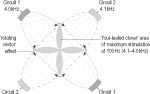
Neurogenic arm (brachial) pain can arise from a variety of causes and become very distressing. Treatment can be problematic, especially if the condition is complicated by the presence of chronic regional pain disorder (CRPD) (see below, ‘Complex regional pain disorder’). A number of work-related conditions are sometimes considered under the umbrella terms cumulative trauma disorder (CTD) or repetitive strain injury (RSI), sometimes also known as repetitive motion disorder (RMD). These include thoracic outlet and carpal tunnel syndromes. Ergonomic aspects of the workplace need to be considered in any treatment programme for CTD, which may result from incorrect and static posture with resultant muscle tension, as well as overuse of particular parts of the body. Musicians may be prone to RSI, for example.178 Exercise, exercises, moving around and relaxation can all be vital. This somewhat controversial diagnosis179 was introduced in the section on peripheral vascular obstruction (see SubCh. 9.6). It is usefully described in Lü Shaojie’s book on nervous system disorders,180 and also in an online article by Alejandro Katz.181 Symptoms include pain and paraesthesiae in the arm and scapula, with the ulnar nerve affected in 90% of cases. Thus ulnar somatosensory evoked potentials (SEPs) are usually abnormal,182 and the middle, fourth and little fingers tend to be most affected.
In a study from Pakistan, local points such as SJ-14 and LI-15 were combined with LI-4, LI-11 and ST-38 distally.186 Joseph Wong suggests using points in the C3–C4 segment for the scalene muscle, HE-1, LU-2 or ST-12 for the brachial plexus and appropriate distal points on the arm.187 Katz applies microcurrent to P-6 and/or SI-3 (negative) connected with anterior scalene muscle TrPs (positive), with LIV-4 and/or ST-41 as secondary points (usually contralateral). He uses surface electrodes positioned over inserted needles. CTENS was found to be helpful for thoracic outlet syndrome in one uncontrolled study.188 Compression syndrome of the lateral cutaneous forearm nerve Meralgia paraesthetica (below) is a recognised disorder. Less well known is the corresponding condition in the arm. Compression of the lateral cutaneous nerve of the forearm, sometimes the result of vigorous exercise, can lead to pain but decreased tactile or pinprick sensation along the radial aspect of the forearm, tenderness to palpation over the nerve where it pierces the deep fascia of the arm (lateral to the bicipital tendon and proximal to the elbow crease) and decreased elbow extension when the arm is fully pronated. Electrical evoked response measurements will confirm the diagnosis. Treatment for this condition has included TENS and ultrasound applied at the tender region of the upper arm.189 Joseph Wong has suggested points such as GB-31, ST-31, ST-32 and SP-12.190 Ulnar entrapment at the elbow is not uncommon, with symptoms of paraesthesiae along the Small Intestine meridian from the elbow towards the wrist and hand. It is likely to be present if light tapping on the cubital groove or SI-8 (with the elbow flexed at 90 degrees) reproduces these sensations.191
Katz recommends stretching exercises to assist recovery. Katz applies microcurrent stimulation at SI-8 (positive) with SI-3 (negative), ipsilaterally (contralaterally, if there is too much local inflammation to treat locally). He uses surface electrodes positioned over inserted needles. Pressure on the median nerve within the carpal tunnel can lead to paraesthesia in the wrist and hand, with muscle wasting and functional impairment if the condition is severe or longstanding. CTS is not uncommon in pregnancy, menopause or associated with rheumatoid arthritis.193 Inflammation within the tunnel may be associated with repetitive hand movements. However, overuse may also lead to other conditions that may be mistaken for CTS, such as shoulder or elbow problems or ulnar entrapment.194 A nerve conduction study will confirm the diagnosis.195,196 Local steroid injections, ultrasound treatment and oral pyridoxine (vitamin B6) have all been used for CTS, with limited evidence that the first of these (and also oral steroids) may be effective, conflicting evidence on ultrasound and the likelihood that B6 is equivalent to placebo, all in the short term. NSAIDs and diuretics do not appear to give much benefit either. Use of a hand brace may be helpful, as may yoga.197 Acute CTS may respond to ultrasound experimentally198 and, although there is some evidence, if limited, that ultrasound may give some symptomatic relief in the long term199 (if treatment is continued for 7 rather than just 2 weeks200), the majority of those given steroid injections are likely to need a repeat within 2–4 months.201 Without treatment, long periods off work and lengthy recuperation may be necessary (half of all employed people with CTS have missed 30 days or more of work202). Given that only 40% of those undergoing surgery regain normal function (although up to 80% may experience considerable pain relief),203 that non-surgical methods do not seem terribly effective and that repeated steroid injections can actually contribute to neuronal degeneration,204 acupuncture may well be an appropriate intervention to try. Indeed, in 1997 the US National Institutes of Health Consensus Development Conference panel considered that there was at least some good evidence for acupuncture’s effectiveness for CTS,205 although some other reviewers do not agree.206
Some helpful prognostic indicators are outlined in the various LA/microcurrent studies by Margaret Naeser and her colleagues (
The Qi Gong machine (QGM) has been used to treat CTS, applied paraspinally at C7–T1, at the elbow (if painful) and locally, for 10–15 minutes at low intensity (if pain increases, the applicator is moved a short distance away from the area treated for about 5 minutes and then reapplied).218 Stretching exercises may be an important part of treating this condition.219 Chiropractic approaches have also been used.220,221,222 Case study 9.11.1 by Ron Sharp details the treatment of bilateral carpal tunnel syndrome.
EA has been combined with glucocorticoid acupoint injection for this condition (using triamcinolone-A or prednisolone)223 and also with Chinese herbal medicine.224 The combination of LA with microcurrent stimulation for carpal tunnel syndrome or repetitive strain injury has been championed by Margaret Naeser225 ( In the single detailed EA study on CTS located, P-6 and P-7 were the points employed Margaret Naeser uses LA at Heart meridian points near the wrist, P-7 and LU-9 (as well as jing Well and baxie points when treating peripheral neuropathy),229 whereas Alejandro Katz applies microcurrent to ~P-6 (positive) locally, with contralateral ST-41 and/or LIV-4 (negative). Whether or not symptoms are reproduced with palpation of ~P-6 after treatment will indicate which of the two contralateral points is likely to give better results. He uses surface electrodes, positioned over inserted needles if the patient is not too sensitive for this.230 Some authors have emphasised treating points in the neck region for CTS, in addition to limb points.231,232,233
When using microcurrent, Margaret Naeser starts treatment with 292 Hz, following this with 0.3 Hz235 LF EA, ‘to tolerance’, was used in the one detailed study located.236 It is surprising that there are not more EA studies on CTS. With a low-power (5 mW) HeNe or 670 nm diode laser suitable for self-treatment at home, lengthy treatments are necessary. For instance, Margaret Naeser recommends a 7 J dose for P-7, which would take around 20 minutes (!), 2–4 J (6–12 minutes) for other painful areas and 1 J (3 minutes) for the other hand points in her protocol. If there is an improvement with these exposure times then shorter treatments can be attempted in later sessions,237 whereas with the higher-power (15 mW) laser used in her clinical study such long exposure times were not needed ( On the basis of Judith Walker’s finding of a sharp increase in 5-hydroxyindoleacetic acid (5HIAA) urinary excretion after 10 sessions of LILT,241 and their own observation that pain level may decrease noticeably after nine sessions, Kenneth Branco and Margaret Naeser recommend at least 15 sessions for CTS with their LA/microcurrent protocol242 or 4–8 weeks for the home treatment version.243 Traction lesions of the brachial plexus, often involving avulsion of nerve roots from the spinal cord (in motor cycle accidents, particularly), may result both in total and irreversible paralysis and in paroxysmal pain that continues indefinitely and responds poorly to medication.244 However, in a minority of cases early reconstructive surgery may be of benefit.245 Surgery has also been used for iatrogenic plexus lesions induced by radiotherapy.246
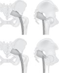
The clunial (cluneal, clunical) nerve, which innervates the skin of the buttock (clunis), has three divisions: the superior, middle and inferior. The former originates with the lateral cutaneous branches of the dorsal rami of L1–L3and the latter two from the dorsal and ventral rami of S1–S3 respectively.253 The medial branch of the superior clunial nerve (SCN) is contained in an osseofibrous tunnel as it passes over the iliac crest just lateral and superior to the posterior superior iliac spine.254 Superior clunial nerve entrapment is an often-overlooked cause of low back pain.255 Pain in the back and buttock, often extending into the leg,256 may begin following back surgery and the formation of adhesions. It can be severe enough to make sitting, walking or standing possible only for short periods.257 Conventionally, this entrapment neuropathy has been treated with steroid injection, thermal ablation or surgery.258 MA has been used for injury of the SCN,259 as have EA260,261 and TEAS.262 EA was found to be more effective than MA in one RCT.263 In one case report of SCS for SCN, TENS gave only partial, temporary relief, whereas the more invasive method was more effective, enabling the patient to return to her normal activities.264 Stimulation of trigger points lateral to L1–L2 may relieve clunial nerve pain.265 Thus huatuojiaji and ashi points have been used for SCN, in conjunction with points such as GB-34 or other distal points. LF EA or TEAS appears to be used more commonly than HF stimulation, although too few studies are available to enable unequivocal recommendation of one rather than the other. The peroneal division of the sciatic nerve passes through the piriformis muscle, rather than under it, in some 10–20% of the population (Fig. 9.11.1). Spasm, contracture or injury of the muscle then compresses the sciatic nerve, leading to symptoms that mimic those of discogenic sciatica, as well as buttock tenderness and pains that sometimes radiate to the lower abdomen and genitals (in some cases even with dyspareunia). Pain may be diffuse, and if the problem is chronic then atrophy of the lateral muscles of the calf may develop. The condition is more common in women than men, in lorry drivers and certain types of athlete.266 It may even be caused by prolonged sitting during extended neurosurgery. In complicated cases, lower lumbar radiculopathy may lead to secondary irritation of the piriformis muscle, while trochanteric bursitis in some cases is due to an underlying piriformis syndrome.267 Further descriptions of the condition can be found in the standard textbooks by David Legge268 and Lü Shaojie.269 Conventional treatment includes physical therapy (ultrasound, TrP injection, stretching and postural correction), steroid injection or surgery.270
Piriformis syndrome has been treated using MA,271 EA272 and a ‘biofrequency spectrum’ device.273 LA has been used for piriformis muscle injury.274 For sciatic pain presumed to be due to piriformis muscle spasm (cramp), Willem Khoe has used ultrasound acupuncture (1 W for 60 seconds) at acupoints such as BL-27–BL-29 and LIV-12, at the greater trochanter and rectally, combined with 2 Hz EA at the L5 huatuojiaji points (15–20 minutes, to tolerance) and vitamin B12 point injection at auricular and hand points for the low back.275 It is unclear which were the most effective ingredients in this cocktail. Other authors have combined EA and acupoint injection for piriformis syndrome,276 for instance using glucocorticoid (triamcinolone-A or prednisolone) injections.277 EA has also been combined with TDP irradiation.278 For non-radicular compression lesions of the sciatic nerve, John Thompson has recommended that TENS electrodes are applied segmentally and proximal to the lesion.279 For sciatic pain presumed to be due to acute piriformis syndrome, Willem Khoe used KI-8, the xi -cleft point of the yinqiao mai (Yin Heel Vessel) extra meridian in his own variant of Yoshio Manaka’s diode cord treatment.280 There are too few studies available to enable generalisations on the most appropriate parameters to use. Both LF and ‘CW’ EA have been utilised ( Nerve compression or injury in the groin can sometimes lead to neuritis in the lateral cutaneous nerve of the thigh, with symptoms in the lower two-thirds of the anterolateral thigh. It may occur during or following pregnancy, or in obese individuals or even as a result of wearing overly tight clothes281 (‘jeans disease’282). This condition has been treated with MA together with acupoint injection,283 with EA together with cupping,284 with electric plum blossom needling combined with moxibustion285 and also with TENS.286 Local stimulation was emphasised in the EA (HF) and plum blossom (LF) studies. Chronic postamputation pain is common, with both central and peripheral mechanisms responsible for its initiation and maintenance. Pain may be experienced in the stump or phantom pain, and may be burning or tingling, or cramping. The former tends to be associated with decreased blood flow in the residual limb and the latter with muscle spasm (cramp).287 To avoid the development of chronic pain, early treatment can be important.288 In one retrospective review of pTENS for chronic pain, it gave useful effects in 65.8% of cases of pain due to amputation.289 In contrast, some surgeons believe that TENS offers no lasting relief of pain following amputation surgery.290 It may be that this difference of opinion is because TENS is more likely to be helpful for milder postamputation pain. Stump pain is sometimes caused by ‘ectopic’ sensory nerve discharges from a neuroma.291,292,293
Ultrasound has been used for stump neuroma pain.299 Electric currents can be measured at regenerating finger stumps in children300 (or whole limb stumps in adult newts301). Regeneration is highly unlikely in human adults, but EAV has been used for amputation pain both as a method of evaluating changes in acupoint electrical potential and for treatment.302 Although such treatment may help pain, there are no reports of it encouraging human limb regeneration, even though theoretically this may be possible. The effect of amputation on the low electrical skin impedance found along meridians has been investigated.303 Both MA and TENS were found useful in one report on stump pain. PEMF, however, sometimes exacerbated it.304 For stump pain, John Thompson recommends that TENS electrodes are positioned paraspinally (in the same dermatome) and actually on the stump.305 TrP injection with a local anaesthetic agent has been used in the treatment of postamputation pain. Contralaterally symmetrical tender points with low electrical skin resistance to an applied 3 V DC signal were selected.306 Roger Coghill recommends magnets locally on the stump itself for stump pain (repeated 15-minute sessions).307 From the studies available, there are no clear patterns on which might be the most effective points to use. Both stump and contralateral stimulation have been tried. From the few studies available on peripheral methods of stimulation for stump pain, it appears that HF stimulation is attempted more frequently than LF EA or TENS. From studies308 and reviews309 of TENS, it does appear that stump pain can respond well to CTENS. However, if it is the case that different sorts of stump pain have different causes, as mentioned above, these should be taken into account. For burning pain and paraesthesiae, parameters likely to improve blood circulation should be used (see SubCh. 9.6), whereas for cramping pain, parameters for muscle spasm (cramp) would be preferable (see SubCh. 9.1, ‘Spasticity in cerebral palsy (CP)’ and SubCh. 9.2, ‘Spasticity’).
Pain in a part of the body that is not there occurs in some 70–80% of patients after amputation, and can be very severe. Surprisingly, it can be congenital as well as acquired.311,312,313 Although it is usually the pain of a phantom limb, phantom pain can also occur after removal of the bladder (or spinal cord injury with deafferentation of the bladder),314 a breast,315,316 or even tonsils.317 Phantom pain, like stump pain, can be burning or tingling or, possibly more often, cramp-like,318 and for the same reasons.319 It should not be confused with non-painful phantom sensation, which may be tactile, kinetic (as if a leg is moving, for example) or even kinaesthetic (a limb may feel as if it is ‘telescoping’ over time). The sense of a phantom limb may be essential for an artificial limb to be usable.320 However, there may be a strong correlation between phantom pain and phantom sensation.321 A phantom limb may be experienced in an awkward position if that is how it was during or just prior to traumatic amputation. It is more likely to be experienced as painful if there was pain at that time,322,323,324 and if the circumstances of the loss were particularly unpleasant.325 However, if it is possible to prevent pain immediately prior to (elective) amputation, for instance with regional anaesthesia,326,327 then phantom pain is less likely to develop afterwards. This may be the result of preventing changes in the primary somatosensory cortex that appear to accompany the development of phantom pain.328 For a similar reason, acupuncture has sometimes been recommended early on in rehabilitation.329 Interestingly, if phantom limb pain is relieved the phantom limb may seem to reposition itself ‘normally’.330 (This has been observed to occur in stages with repeated acupuncture treatment, for example.331) Whether general or regional anaesthesia is used during amputation appears to have no bearing on whether phantom pain develops or not.332 Although from experimental studies it would appear that pain relief immediately postamputation is not likely to prevent the subsequent development of phantom pain, continuing early acupuncture treatment for some weeks has been recommended.333 Further surgery is unlikely to be helpful.334 As with many forms of chronic neurogenic pain, complete relief of pain may not be a realistic objective, although pain reduction may well be possible. The level of pain experienced may be related to stress, anxiety, exhaustion, and depression,335 and treatment regimens may need to take these and other contributory factors into account. Acupuncture, both MA and EA, has been used for phantom limb pain336,337 (
In addition to phantom limb pain, TENS has also been used for phantom bladder354 and phantom breast pain. In the latter case, when tolerance developed to treatment (that also incorporated auricular acupuncture and medication), metastases were subsequently detected.355 Electrical stimulation (‘faradisation’) of the brachial plexus was early on found by Weir Mitchell (see below, ‘1863’) to ‘resurrect’ a phantom hand that had been missing for 25 years.356 Both vigorous acupuncture357 and TENS358 have been reported as eliciting or exaggerating phantom limb pain (TENS applied to the stump could also revive a phantom that had been successfully suppressed).
Gary Null has suggested that improved circulation in response to magnetic field application may also benefit phantom pain.360 Timo Töysä has found that magnets even well away from the affected limb may be helpful for phantom pain (he interprets this as supporting the meridian hypothesis).361 Pulsed magnetic fields have also been applied at acupoints for phantom pain362 ( SCS has been used for phantom limb pain, although with unpleasant side-effects in some cases.363 Focusing attention on phantom limb pain will affect it.364 Phantom limb pain may thus respond to EMG biofeedback,365 which offers the interesting prospect of combining such methods with treatments such as EA that are likely to affect muscle activity. In one case report, the patient’s preference was for MA over both EA and TENS.366 In another, the combination of acupuncture and TENS was the only treatment that enabled the patient to wear his forearm prosthesis with a degree of comfort.367 In one uncontrolled study, EA gave better results than TENS; the best results were found with their combination.368 EA (at scalp points) has also been combined with MA and moxibustion for phantom pain.369,370 From the acupuncture-based studies in the database ( Phantom limb pain is liable to be more severe if the stump itself is colder.371 Thus Jacqueline Filshie has suggested that acupoints be used that are likely to block sympathetic activation.372 In the case of phantom breast she has reported using both paraspinal (T1/T2) and suprascapular TrPs to good effect (the latter to reduce muscle spasm (cramp)). In one case report, MA was effective only at one very specific point.373 With conventional TENS, the peripheral nerve trunks are usually stimulated. However, if these are absent, as in the phantom limb, other strategies have to be adopted. David Bowsher, for example, suggested that the spinal cord itself should be stimulated at the segmental level involved.374 Other TENS electrode placements have included TrPs, acupoints, segmentally or contralaterally, over related peripheral nerves, or the stump itself. John Thompson, for instance, has recommended positioning electrodes as for stump pain (paraspinally and on the stump).375 Gessler and Struppler found that TENS over the nerve supplying extensor muscles of the phantom was particularly helpful, giving the impression of cramplike flexures ‘opening’ and relaxing.376,377 Laitinen and others obtained good results with contralateral stimulation.378,379 Given that local stimulation may exaggerate phantom pain, contralateral treatment should perhaps be attempted first, as mentioned. This approach has been tried using press needles at contralateral points, for example,380 as well as with local anaesthetic injections.381 However, TrP anaesthetic block does not always give good results.382 When to use what – parameters for phantom pain EA studies have predominantly employed LF stimulation, ‘to tolerance’, or at ‘bearable’ intensity. In the TENS studies, a wide range of parameters has been used. CTENS is sometimes recommended,383 but is not always helpful.384 LF burst TENS is another possibility,385 although it was less effective than CTENS in one study where electrodes were positioned to stimulate phantom extensor muscle efferents.386 ALTENS has been used contralaterally.387 Given the likely involvement of an affective component and spinal segmental mechanisms in the development of phantom pain, MA and low-intensity HF EA or TENS have been suggested as appropriate parameters to use both preoperatively (for 3 days when possible) and following amputation.388 Rather as with CRPD, a gentle approach emphasising a sense of inner stillness has been recommended rather than treatment heroics that could reinforce the initial trauma.389 As with stump pain, parameters should be selected in accordance with whether the phantom pain is more burning or cramping (see above, ‘burning pain and paraesthesiae’). Peripheral sensory neuropathy may accompany endocrine conditions such as diabetes or hypothyroidism, or may result from infectious diseases such as leprosy, Lyme disease or HIV, some nutritional deficiencies (thiamine, folic acid, vitamin B12) or ingestion of toxic chemicals (including long-term use of alcohol or some medication). Unexplained sensory neuropathy may be associated with cancer that is only diagnosed many months later – the result of nerve compression from a growing tumour. Mixed sensory and motor neuropathies occur in uraemia and Guillain–Barré syndrome, as well as in most types of Charcot–Marie–Tooth syndrome,390 the commonest inherited peripheral polyneuropathy.391 (In most variants of this condition as in multiple sclerosis it is the myelinated rather than unmyelinated fibres that are most affected,392 although one type involves axonal degeneration alone.393) When acute, the symptoms of axonal polyneuropathy associated with excess alcohol consumption in conjunction with poor nutrition may resemble those of Guillain–Barré syndrome (see SubCh. 9.3), but without involvement of the immune system.394 In TCM terms, peripheral neuropathy is usually considered under the heading of wei bi, or wei syndrome (sometimes clumsily but expressively translated as ‘wilting-impediment syndrome’).395 Substance P (SP) may be low in the cerebrospinal fluid (CSF) of patients with peripheral neuropathy (but may be raised in those with arachnoiditis, see SubCh. 6.2, ‘peripheral neuropathy’). Acupuncture has been suggested for the neuritic pain of leprosy,396,397 although I do not know whether it has actually been used for this to any extent.
EA, Chinese herbal medicine and acupoint injection have been used together for drug-induced multiple radicular neuritis.412 Methylcobalamin (mecobalamin), the neurologically active form of vitamin B12, may be beneficial for both peripheral and autonomic neuropathy.413,414 A combination of peripheral electrostimulation with this approach should be investigated to see whether results could be improved further. A case of ulnar neuropathy following carpal tunnel surgery responded well to a combination of MA with high potency B-complex vitamins in my own practice. For drug-induced neuropathy, the most commonly used points appear to be ST-36 for the lower limb and LI-11 for the upper limb, although of course usually not treated in isolation. Electrode placements in the TENS/TEAS studies of peripheral neuropathy include acupoints, and proximal to or over painful areas. With a two-output TENS machine, Gordon Gadsby and Michael Flowerdew suggest that one pair of electrodes can be positioned at the site of neuropathic pain and the other pair bilaterally at ST-36.415 One interesting approach tried for peripheral neuropathy combined acupuncture with home use of cathodal TEAS at ST-36 for the leg, or LI-11 for the arm, the return electrode (anode) being placed in a waterbath in which the whole of the affected foot or hand was also immersed.416 The Liss Pain Suppressor device was used in this case report, in which a soothing sensation was experienced rather than the tingling felt with conventional TENS ( Corresponding to her protocol for CTS and neuropathic hand problems ( When to use what – parameters for peripheral neuropathy ALTENS has been found useful for experimental neuropathy in rats (see SubCh. 6.4, ‘WDR activity’). CTENS or TLEA, especially if not charge balanced, may not be suited to peripheral neuropathy with hyperaesthesia or serious sensory loss (see Ch. 12 , ‘TLEA, especially’). In the first instance local treatment may serve only to exacerbate pain, whereas in the second skin damage could occur without the patient’s awareness if treatment is continued for long periods. It has been suggested, on the one hand, that stimulation at > 50 Hz may not be appropriate for peripheral neuropathy if denervation is a factor (see too Ch. 12). On the other, experimental research indicates that HF may more effectively reduce neuronal hyperexcitability than LF stimulation (see SubCh. 6.4, ‘way: more’). Furthermore, increased blood supply to the (sciatic) nerve, which presumably would be beneficial in cases of neuropathy, may occur only in response to HF and not LF EA.419 However, evidence from clinical studies on the effectiveness of CTENS for peripheral neuropathy, for example, is limited.420,421,422 Clinically, moderate intensity DD or LF EA has been recommended for polyneuropathy,423 although, as mentioned (see SubCh. 9.10, ‘parameters for nociceptive and neurogenic pain’), some practitioners do prefer to use CTENS/TLEA. From the studies in the database ( Metabolic neuropathy may result in part from oxidative and nitrative stress (involving the vasodilator nitric oxide, NO). Adjunctive treatment with antioxidants such as alpha lipoic acid may therefore be beneficial.424 Peripheral neuropathy is the commonest complication of diabetes mellitus.425 It may be evident in gait and balance abnormalities (postural dizziness) even before diabetes is diagnosed,426 and may already be evident in children and adolescents with poor metabolic control of their condition.427 In older diabetics, peripheral neuropathy can also be associated with peripheral ischaemia due to vascular disease. In fact, chronic peripheral arterial occlusive disease may itself cause axonal polyneuropathy,428 and it can sometimes be difficult to tell whether pain is due more to diabetes or to poor circulation (as in some other conditions where both may be involved such as erythromelalgia ( It is important to remember that diabetic autonomic neuropathy can cause dysfunction or actual damage in many internal organ systems – cardiovascular (e.g. tachycardia), gastrointestinal (e.g. gastroparesis, diarrhoea), genitourinary (e.g. neurogenic bladder), adrenal and ocular (pupillary abnormalities).433 Also, with reduced afferent input from limbs affected by peripheral neuropathy, neuropathic changes can occur centrally as well. Thus afferent stimulation can be an essential part of treatment.434 Acupuncture has been used for both autonomic and peripheral diabetic neuropathy.
For peripheral diabetic neuropathy, results with EA were superior to those of MA (mock EA) in one crossover RCT (see Ch. 8, ‘diabetic neuropathy’). EA was also superior to medication in other controlled studies.438,439 Improvement was found to peak after about 6–8 sessions by Romanian researchers.440
Strong (painful) TENS may well elicit axon reflex vasodilatation when applied to the top (dorsum) of the foot. However, this response may be absent or reduced in those with diabetic neuropathy (this in itself may be useful diagnostically,444 (see SubCh. 6.3, ‘axon reflex’). Various forms of TENS have been used to screen for peripheral neuropathy.445
In some interesting research by Constantin Ionescu-Tîrgovişte and Romanian colleagues, electrical potentials measured through needles inserted ~1 cm into acupoints were reduced, and sensory threshold increased, both in diabetics compared with non-diabetics448 and in diabetics with neuropathy compared with diabetics without neuropathy.449 Norman Shealy reports good results for diabetic neuropathy by combining CES (1 mA for 1 hour daily, using the Liss device) with MRT using experimental GigaTENS. Each of the 12 acupoints in his ‘ring of fire’ protocol was irradiated for 3 minutes daily, using 52–78 GHz at 1 µW, for two 5-day courses separated by an interval of 2 days, and then 10 once-weekly treatments. Progesterone cream was also applied.450 As with other forms of neuropathy, the most frequently used point is probably ST-36. Other Stomach and Gall Bladder meridian points are often used in addition, perhaps less commonly with points on the Bladder, Liver, Large Intestine and Spleen meridians. TENS studies usually focus on treating the leg and foot directly, perhaps with paraspinal lumbar electrodes in addition. An electrode sock, stimulating the whole lower leg non-specifically but particularly those regions where electrical skin resistance is low, is an interesting innovation451 (glove electrodes are also available). It appears that LF stimulation is used more frequently than HF in the treatment of diabetic neuropathy. DD EA and HF TENS have also been utilised. There is little information available on the current intensities employed. HIV-related peripheral neuropathy Neither MA nor amitriptyline was found to be superior to placebo in one study on HIV-related peripheral neuropathy.452 However, both EA453 and pTENS454 were helpful in single case reports on this condition.
There is little consistency in point selection for neuropathy purely due to HIV/AIDS (without further iatrogenic neuropathy), and little information on appropriate stimulation parameters. Unpleasant (not usually painful) sensations in the legs, often disturbing sleep and relieved by movement, may be associated with peripheral neuropathy455 as well as impaired peripheral circulation (see SubCh. 9.6). This ‘restless legs syndrome’ has been treated with MA,456 EA457 and TENS458,459 ( BL-56, ST-36 and SP-10 have been used, among other points, for restless legs syndrome, as well as interrupted, LF and HF stimulation. As yet there are too few studies to hazard a guess as to what approach is optimal for this problem, which like so many is multifactorial in origin.
Radiculopathy-related pain Radiculopathy-related pain is due to a combination of mechanical and biochemical events. Compression of the nerve root creates oedema, leading to intraneural inflammation (neuritis) and hypersensitivity.463 Symptoms may be sensory (hyperalgesia, allodynia), autonomic (vasoconstriction, excessive sweating on movement or needling, ‘goose bumps’, peau d’orange, dermatomal hair loss), or motor (muscle shortening, reduced range of movement, thickening of tendons where they attach to bone).464 Thus radiculopathy may alter both motor and sensory function. Neurologically, pathology is confirmed by needle electromyography (EMG) and somatosensory evoked potentials (SEPs) respectively,465 although abnormal EMG findings may occur in those without symptoms and should not be considered in isolation.466 Motor tract stimulation with assessment of evoked potentials from several muscles simultaneously can also be useful for diagnosing motor radicular lesions.467 When needling paraspinal muscles for the purposes of EMG evaluation, patients may report reproduction of their symptoms (pain or paraesthesia),468 suggesting inadvertent TrP treatment while (or rather than?) confirming the level of the root lesion involved. Indeed, for many medical acupuncturists, dry needling of myofascial TrPs is a key component of successful treatment for radiculopathic pain and related symptoms. Chan Gunn, for example, claims that all myofascial pain conditions are, in origin, radiculopathies that cause concurrent muscle shortening that in turn perpetuates the radiculopathy by increasing disc compression. For Gunn, even peripheral lesions (as in carpal tunnel syndrome) and osteoarthritis may be secondary to nerve root entrapment. Thus relaxation of paraspinal muscles is a key to successful treatment.469 Peter Baldry, in contrast, considers that only around 10% of those with myofascial TrPs require deep dry needling to release nerve root compression.470 Of course, there are causes to back and neck pain other than radiculopathy,471 and differentiating whether these are ‘purely’ muscular or are due to nerve root compression or other nerve entrapment, let alone the aetiology of such entrapment, is by no means an easy task. As is well known, much chronic back pain remains a mystery, for all the hi tech scans and invasive interventions now available,472,473 although careful evaluation obviously helps to reduce the proportion of those who fail to improve with standard care.474,475 Here low back pain and sciatica will be considered. Neck conditions are covered later on in this chapter (see below, ‘Neck pain’). Back pain can be considered under various headings, based on whether or not nerve compression or entrapment is an important factor ( Sciatica is usually due to pressure on a nerve root and ensuing inflammation (neuritis), although piriformis syndrome (see above, ‘Piriformis syndrome’) and some forms of neuropathy may give similar symptoms.476 Very occasionally, conjoined nerve roots may lead to low back pain and sciatica, even without impingement on the nerve.477 A nerve root problem is most likely if flexion is not problematic, but extension and side bending towards the problem side increases pain in the affected extremity.478 L4–L5 and S1–S3 nerve roots are those most commonly affected.479 Thus L4–L5 lumbar disc herniation is one possible cause of sciatic pain. As Daniel Traum has pointed out, if L3–L4 nerve roots are affected then pain is likely to radiate to the sacroiliac joints, hips, posterolateral thighs and fronts of legs, with anteromedial numbness, weakness of knee extension and a decreased knee reflex. If L4–L5 nerve roots are involved, pain will radiate in much the same way (but down the posterolateral rather than anterior lower leg). Numbness in this case will be lateral, perhaps extending into the dorsum of foot and big toe with weakened foot dorsiflexion but reflexes normal. L5–S1 and S1 nerve root involvement will result in a similar pattern of pain radiation with numbness in the lateral leg, calf, foot and heel, as well as in the sole. Plantarflexion may be weak and the ankle jerk reflex reduced or absent.480 As a rough guide, L3–L4 symptoms occur along the Stomach meridian, L4–L5 symptoms along the Gall Bladder meridian and L5–S2 symptoms along the Bladder meridian.481 However, radicular symptoms usually take around 10 years, on average, to show up after an initial episode of acute traumatic low back pain,482 and disc protrusion may be present in up to 50% of those aged over 40 who do not experience back or leg pain at all.483 Bilateral sciatica, with ‘saddle’ anaesthesia, lower-extremity weakness, loss of bladder control and impotence, is likely to indicate cauda equina syndrome rather than a radicular problem. Interestingly, patients with sciatica who do not recover well from surgery for herniated lumbar disc may well have a smaller-diameter spinal canal as well, and so a degree of stenosis484 (although not as severe as in cauda equina syndrome, when the nerve roots descending within the canal are themselves compressed). Conventional treatments for nerve root problems include disc surgery, radiofrequency ablation of the dorsal root ganglion of the implicated nerve and epidural steroids.485 However, repeat surgery is often unhelpful,486 epidural steroids give only limited and unsustained relief,487 sciatica is generally considered amenable to electrostimulation488 and pain due to disc herniation is well documented as responding to non-invasive treatment in many cases.489,490 For more resistant problems, SCS has been used – for chronic pain due to cervical radicular damage491 or ‘battered root syndrome’ following spinal surgery.492 In general, reversible radicular pain due to entrapment is more likely to respond to treatments such as acupuncture than established nerve damage (neuropathy).493 For this reason, as indeed with surgery,494 better results are likely if treatment is started within 3 months of sciatic pain onset. Sensory deficits are more likely to persist than motor problems.495 Chen Ke-zheng has written one of his useful brief reviews of various acupuncture methods on the treatment of sciatica.496
In Russian studies, pain reduction was associated with improved alpha motoneuron reflexes, more on the healthy than on the affected side (using plum blossom needling together with EA),503 and also with improved local and systemic circulation (with subdermal needles retained for 2–3 weeks).504 MA (combined with electrotherapy) also enhanced electrical excitability of muscle, in patients with sensory and motor disturbance attributable to lumbosacral radiculitis.505 For an acute exacerbation of long-term sciatica, MA together with antidepressant medication both extended short-term pain relief and reduced the number of days that treatment was needed, compared with MA (and placebo medication).506
Case study 9.11.2 is an example of the use of electroacupuncture for acute back pain.
Although paraesthesiae should be elicited by TENS in the affected area, the pain itself may be reproduced when TENS is applied; this may occur, for instance, with sciatic radicular pain.516 In one mixed study, CTENS was found to be less effective for radicular than for purely musculoskeletal pain.517 In keeping with this, TENS may be more useful when pain is relatively localised,518 but less so for sciatic or radiating pain519,520 or if several nerve roots are involved.521 It will also be less effective if there are signs of denervation522 (spinal nerve root compression, with ‘loss of function’523,524).
‘Microwave acupuncture’ has been used for sciatica,526,527 sometimes combining microwave stimulation with needling,528,529 or with other physical therapy methods.530,531
Iontophoresis at acupoints has been used for lumbar pain (including that due to disc prolapse), with results superior to those of conventional medication.539 However, in a study comparing MA and iontophoresis, neither was effective in cases of root compression associated with lumbar osteochondrosis.540 Acupoint/ashi point injection is not infrequently used for sciatica.541,542,543 Simple saline injection (not necessarily at traditional acupoints) has also been utilised.544 Stretching and gentle movement to relax muscle paraspinal muscles can be important for sciatica sufferers.545 Inactivity may be counterproductive. Although sciatica associated with lumbar disc herniation (protrusion) may respond well to acupuncture, if the protrusion is large or both spinal cord and nerve root are compressed then acupuncture is less likely to be beneficial and surgery may be essential.546 However, even if surgery is necessary to avoid injury to the nerves and muscle atrophy in the lower limb, acupuncture may still be helpful to aid recovery and reduce pain postoperatively.547 EA was better than MA for sciatic pain in one controlled study,548 and better than simple retained needling for intervertebral disc protrusion.549 EA was also preferable to MA for sciatica in other reports,550 for instance using periosteal stimulation (with both MA and EA).551 However, in another CT, MA (based on TCM syndrome differentiation) gave better results than EA for sciatica.552,553 Pekka Pöntinen has warned that exacerbation due to hyperstimulation is more likely with EA.554 Similarly, Sin Yoke Min has cautioned that some patients can become sensitised to EA, which should therefore not be used at every session.555 Perhaps for this reason, Wu Jinwei has recommended that only gentle EA be used in some patients.556 MA was superior to acupoint injection of Chinese herbal medication in one report on sciatica.557 EA and TEAS were both found effective for sciatica in one comparative study.558 However, in low back pain due to degenerative disc disease EA (‘PENS’) at non-acupoints gave better results than paraspinal TENS.559 Also, following surgery for spinal osteochondrosis, EA was more helpful for pain than TENS with the same sinusoidal waveform and frequency, although both methods were effective for associated motor deficit.560 MA was more effective than EA for sciatica in one comparative study in which syndrome differentiation was emphasised.561 In one RCT, LA gave better results than EA and ultrasound acupoint stimulation in discogenic lumbosacral radiculopathies.562 Microwave acupuncture was superior to MA in its effects in one study.563 EA for sciatica has been combined with various treatments, including TENS,564 the shendeng ‘sacred lamp’565 or other multifrequency devices,566 massage567 and acupoint injection.568,569,570 The latter combination was found to improve effectiveness,571,572 as was the addition of both acupoint injection and warm needling573 or a multifrequency lamp574 to simple EA. EA has also been combined with both infrared heat and point injection.575 EA with warm needling gave better results than just EA.576 Similarly, TDP irradiation with either MA577 or EA578 gives better results than MA alone. In contrast, the addition of acupuncture to ‘biofrequency spectrum’ treatment did not improve results in another report.579 The combination of EA and moxibustion with massage gave significantly better results than their combination with traction in one study where ultrasound scanning of prolapsed intervertebral discs was used to confirm these improvements objectively.580 When combined with EA and herbal ‘steaming’ the results of traction were improved.581 EA with medication applied to the skin, although perhaps more effective than EA alone, was not superior to combined massage and traction.582 As with cervicobrachialgia, combining MA with intradermally implanted needles improves results in radicular lumbar pain. Not only are benefits maintained for longer, but the combination has a greater effect on peripheral circulation as well.583 In acute episodes, combining MA with antidepressant medication (amitriptyline or pyrazidol) also increases the duration of analgesia after treatment and reduced the number of treatments required.584 MA is often combined with massage for sciatica,585,586 or with cupping.587 In one report, it was combined with TENS, traction and HF electric field treatment.588 It is important to note that acupuncture (MA, EA and warm needling) gave better results in one study when combined with conventional conservative treatment.589 Also, MA together with Chinese herbal medicine (CHM) may give results little inferior to those of steroid nerve block and better than those obtained with local anaesthetic injection.590 Huatuojiaji591 and ashi592 points can be important when treating radiculopathic pain such as sciatica, but strong local stimulation at hypersensitive points, or at many points, should be avoided at least initially. Bladder meridian points (BL-11, BL-23, BL-36, BL-40, BL-54, BL-57, BL-60, BL-62), Kidney points (KI-3, KI-7), Gall Bladder points (GB-30, GB-34, GB-40) and points such as ST-36, ST-44, SP-6, Du-2, Du-3, Du-4, Du-15 and Du-20 may all be relevant.593 From the studies in the database ( With TENS, long electrodes can be positioned paraspinally to span several nerve roots (for instance, L1–L5 or L3–S2 for sciatica).596 For sciatic pain, one microcurrent advocate positions electrodes anterior and posterior to the affected nerve root,597 whereas a manufacturer of microcurrent devices recommends positive stimulation over the nerve root (at L4 or L5), the negative electrode being positioned at BL-40.598 With the Likon device, stimulation is suggested locally, or distally over the Bladder meridian.599 With LILT, Margaret Naeser and Xiu-Bing Wei suggest directing the beam at the nerve root site from three or four different directions, and then adding LA at points such as LI-4 (for cervical radiculopathy) or BL-60 or BL-67 (for lumbar radiculopathy).600 In one MA study of sciatica, the combination of a single body point with scalp stimulation gave better results than with body points alone.601 When to use what – parameters for radicular pain DD mode has been recommended for muscle spasm (cramp) in neurological conditions,602 and so could be considered for radiculopathic pain. It has been used in a number of EA studies on sciatica and lumbar radicular pain, for example in database sections ( In one report of EA for the sequelae of polio, stroke and sciatica, the strongest stimulation was required for the first of these and the least strong for sciatica.603 However, the author of one MA/point injection study noted better results for sciatica with stronger stimulation.604 In the main, although obtaining deqi is important and the usual phrase ‘to tolerance’ appears frequently in the EA sciatica and low back pain studies, my (biased!) impression is that high-intensity stimulation is not emphasised unduly for these conditions. Indeed, some authors have emphasised that treatment should be gentle, at least in some cases.605 The authors of one retrospective mixed pain CTENS study observed that, whereas results were comparatively poor for radicular pain, they did improve with higher intensity stimulation.606 An interesting comment was made by Tian Deming, author of one uncontrolled study on sciatica, who stated that HF EA can be used for patients in robust health, but that LF EA should be used if not. There are certainly more LF than HF EA studies in the database In an early trial comparing ALTENS and CTENS for chronic low back and leg pain, analgesia was short lived with both modes.609 More patients found CTENS to be helpful, possibly because ALTENS was too strong for comfort. In a very small (N = 8) study comparing CTENS, burst and modulated TENS, three out of four sciatica patients responded best to burst TENS. At 2 months, two still benefited.610 However, both motor level burst TENS and CTENS were found to give prolonged analgesia in one Russian study on lumbar and sciatic pain due to spinal osteochondritis.611 In one EA (‘PENS’) study on degenerative disc disease, 30 minutes of stimulation gave better results than 15 minutes.612 When using microcurrent applied through pads, 20 minutes of stimulation has been recommended as a minimum treatment duration.613 As for polarity, as mentioned (see SubCh. 9.10) ‘positive probe proximal’ may be appropriate, the distal probe being positioned at a point in the area of referred pain in the leg. As pointed out above, it is often considered that radiating pain is more difficult to treat than purely localised back (or neck) pain. Whether non-charge-balanced or DC stimulation would improve results for radiating pain compared with biphasic charge-balanced treatment has not been adequately researched. Needle depth may need to be adjusted according to whether sciatica is acute or chronic. Apart from Gunn, a number of authors consider deep needling to be important.614,615 In one MA RCT, however, there was little difference in outcome whether GB-30 was needled to a depth of 0.5–1 cun or 2–3 cun.616 Case study 9.11.3 details the use of automated methods of intramuscular stimulation in a case of low-back pathology.
Complex regional pain disorder Complex regional pain disorder (CRPD) has been described as a devastating disease.617 It was first recognised as a clinical entity in 1863 by Silas Weir Mitchell, who observed the burning pain caused by gunshot or other nerve injuries sustained in the American Civil War. Today, in the US alone, some five to six million people are estimated as suffering from CRPD.618 The pain, which may affect one or more extremities, can become intense and quite disproportionate to the underlying pathology.619 CRPD may follow even slight injury, which means it is often underrecognised particularly in children620,621 (however, one perhaps unsympathetic reviewer considers it is over diagnosed!622). Estimates vary widely on the proportion of sufferers likely to recover spontaneously. CRPD may come in two guises,623 which are not always clearly distinct. Type I corresponds to what used to be called reflex sympathetic dystrophy (RSD), and is more likely to be diffuse, without following any dermatomal or other logical distribution.624,625 CRPD type II tends to have a more clearly somatic origin, and corresponds to causalgia (pain and hyperalgesia, confined to the distribution of an injured peripheral nerve626). CRPD can accompany repetitive strain injury (RSI), peripheral neuropathy or entrapment/compression syndromes, or follow burn or fracture injury, surgery (spinal627 or mastectomy,628 for example) or other insult to an organ (for instance, after myocardial infarction or as ‘shoulder–hand syndrome’ following stroke,629 see SubCh. 9.2). It has even been suggested that interstitial cystitis may be a form of reflex sympathetic dystrophy.630 Not only sensory but also sympathetic nerves can be involved (hence the earlier name ‘reflex sympathetic dystrophy’, RSD). Various central and peripheral mechanisms may be implicated.631,632 Following hand surgery, for instance, it has been suggested that CRPD type II (sometimes termed ‘algodystrophy’) may correspond to an exaggeration of normal post-traumatic inflammatory responses rather than sympathetic damage, and can be prevented by minimising postoperative inflammation, increasing venous return (especially by avoiding constrictive dressings or casts), controlling post-traumatic pain and promoting early active mobilisation.[633] If this is so, it may provide a model for treating some non-iatrogenic CRPD if the initial injury does not go ignored. In the first stage of CRPD (type I), sympathetically maintained pain is accompanied by sensory changes, allodynia, hyperalgesia, non-pitting oedema and sudomotor and vasomotor changes (the affected extremity is likely to be warm, dry and red or slightly cyanotic). However, sympathetic symptoms such as local sweating or changes in skin temperature and colour may not be present,634 and the degree of hyperalgesia or allodynia experienced may not correlate with the intensity of spontaneous pain.635 Even at this early stage, there may be localised changes in hair growth and nail texture.636 Stage 1 usually lasts for 1–3 months, but may persist indefinitely.637 In the second stage, the symptom picture widens and may include inflammatory changes in the skin, neurodermatitis, bruising, tremor, joint swelling, insomnia due to night pain, and emotional disturbances. The skin is likely to become shiny, pale, cool and clammy.638 Motor function can be impaired.639 Hair growth now diminishes, and there may be changes in bone structure.640 In some stage 2 CRPD, serum noradrenaline may be lower on the painful side than on the healthy side.641 In the third stage there may be spontaneous skin ulceration, infection, muscle atrophy, flexion deformities and osteoporosis, as well as the depression that may accompany any form of chronic pain.642 Treatment for CRPD has to be intensive and multimodal. Ted Priebe, on whose useful account of CRPD much of the above description was based, recommends that exercises should be given along with acupuncture on the same or alternate days. Other practitioners precede occupational or physical therapy with sympathetic blockade, for example: stellate ganglion blocks for upper limb problems and lumbar block for lower limb symptoms.643,644 At least six sympathetic blocks may be needed before there is any response (as with acupuncture the effect can be cumulative). It is important that exercise should not be forced or painful (‘only gain with no pain’ rather than ‘no pain, no gain’645). Surgery is likely to exacerbate the problem.646 As with PHN, treatment has to be started in the early stages of the condition (certainly within 6 months647) for best results. Response may be slow, and once the condition has become chronic, it may become intractable.648 (One estimate is that this occurs in around 40% of CRPD patients,649 another in some 10%.650) If it does respond, treatment may need to be repeated at intervals indefinitely.651 Spontaneous remission is rare.652 However, in children full recovery is much more likely than in adults.653,654
Theoretically acupuncture, which can reduce both sympathetic overactivity and pain, should be helpful for CRPD.659 Furthermore, in experimental studies both MA and EA have been shown to reduce some components of acute neurogenic inflammation,660 and thus may possibly be of benefit as a preventive measure in the very early stages of CRPD. Perhaps for this reason, various types of acupuncture (such as magnets or moxibustion used at acupoints) have been found helpful by children with CRPD (their parents were less convinced).661 In general, however, clinical results with both acupuncture and TENS for CRPD have been described as ‘not very impressive’.662 Thus Joan Hester has commented that whereas acupuncture may be helpful in early stages of the disease it is not as effective as sympathetic blockade and should be used with caution in patients with hyperpathia (hyperalgesia or allodynia or both, possibly prolonged).663 In contrast, in some cases Dharma Singh Khalsa has found acupuncture useful in later stages of the condition when blocks are no longer effective.664 Also, EA was more effective than MA in one study of poststroke shoulder–arm syndrome (not CRPD proper?)665 (
The localised cooling that can sometimes accompany CRPD may be improved by TENS.677 In one quite large retrospective study, TENS (15–25 Hz, modulated) used for 22 hours each day made a considerable contribution to successful rehabilitation not only in terms of pain control, but also for vasomotor symptoms. It was noted that during the first 2 weeks of treatment symptoms returned rapidly if TENS was discontinued, but that after 2–4 months the unit could be dispensed with for several days before symptoms returned.678 Unusually, TENS has been used for CRPD following mastectomy.679
Other electrotherapy methods that have been investigated for CRPD include high-voltage pulsed galvanic (HVPG) and athermal pulsed shortwave (Diapulse) ( Both skin temperature and conductance measurements have been used to monitor changes in CRPD with ryodoraku stimulation.687 From the studies in the database ( For lower-limb CRPD, Alejandro Katz has used microcurrent at ST-36 and the bafeng points between the toes ipsilaterally, or contralaterally for a few treatments if the condition is too painful initially. He positions surface electrodes over the needles.688 In a useful overview of CRPD treatment from a TCM perspective, John Stebbins recommends MA, followed with microcurrent stimulation, at points such as SJ-5, GB-34, LI-11, ST-36, ST-40, SP-6, SP-9, baxie and bafeng, together with points on the affected meridians both proximal and distal to the affected area.689 However, when using TENS, Ronald Melzack and Patrick Wall found that stimulating proximal to an area of causalgia could markedly reduce hyperaesthesia, whereas stimulation distal to the area could aggravate it.690 Furthermore, TENS (or other stimulation) applied to the affected limb/area will probably feel stronger and be tolerated less easily than on the unaffected side, and may lead to uncomfortable spreading sensations that tend to outlast the stimulus.691 Contralateral treatment may thus be advisable.692 In contrast, ipsilateral paravertebral (C6–T3) and anterior neck electrode positioning to ‘sandwich’ the stellate ganglion was found helpful for upper-limb CRPD in one small Italian study.693 There is no reason why non-local auricular EA could not be tried with CRPD. Auricular EA was more effective than MA in one study of acute neurogenic oedema (see SubCh. 6.3, ‘reduced neurogenic’), and ear points have been stimulated in several studies on CRPD. When to use what – parameters for CRPD From the studies in the database ( In animal experiments, deep needling may be more effective than superficial needling for acute neurogenic inflammatory oedema (see SubCh. 6.1, ‘ST-36/GB-30 MA’). However, very strong EA (70 mA!) may aggravate it, although EA at a more clinically appropriate CRPD is a chronic neurogenic condition, and as such may well be exacerbated by strong treatment local or distal to the problem area (see Ch. 12, ‘CRPD’). Thus Christer Carlsson advises against deep needling for CRPD.694 Katz similarly recommends superficial needle insertion and subthreshold stimulation only.695 CTENS is sometimes recommended as appropriate for CRPD.696 30 Hz TENS, ‘to tolerance’, was used in the Italian study on stellate ganglion stimulation mentioned above.697 In a single case report of TENS for CRPD in a young child, 50 Hz (3.5 mA) was eventually found to give better pain relief than 90 Hz (2.5 mA), used initially.698 Extreme temperatures (ice packs or radiant heat) should be avoided.699 The overall message would appear to be that gentle stimulation is most appropriate for CRPD, and that it should be started as early as possible. Treatment may need to be prolonged (see above, ‘Response may be slow’). Central pain is defined by the International Association for the Study of Pain as pain initiated or caused by a primary lesion or dysfunction in the central nervous system.700 There are several reviews of the various central pain syndromes and their treatment.701,702 In an experimental study, EA (and MA) at SI-17 had an anti-inflammatory effect on neurogenic inflammation of the brain’s dura mater greater than that of prednisolone, although this was less than that of saline acupoint injection (see SubCh. 6.1, ‘dura mater’).
5-hydroxytryptophan (5HTP) the serotonin precursor has been investigated for its effects on central and deafferentiation pain,704 with potential implications for its combination with electrostimulation methods. The usual image we have of the stroke victim is of someone with hemiplegia or at least a degree of motor impairment. However, sensory pathways and areas in the brain can be affected as well in the so-called ‘thalamic syndrome’, leading to pain that is sometimes very severe705 but often begins only some months after the stroke itself. This central poststroke pain occurs in up to 8% of patients. It is usually burning and is accompanied by sensory deficit, as well as by allodynia (in around half of patients). As with so many neurogenic pains, it may be reduced by adrenergically active antidepressants such as amitriptyline, but treatment must be started early to take effect.706 Perhaps because motor impairment is so much more visible than central pain, there is a relative paucity of studies on electrotherapy for the latter. Indeed, some authorities consider it unlikely to respond to peripheral treatment methods,707 with stimulation of the brain itself being used instead as a last resort (deep, to the thalamus or central gyrus, or more recently motor cortex stimulation,708 sometimes via extradural electrodes709). Frequent technical and clinical monitoring is necessary in such cases.710
TEAS (at LI-4 and ST-36),712 TENS713,714 and SCS have all been used for central poststroke pain.715 (Indeed, the mechanism of EA at paraspinal points is probably similar to that of the more invasive method of SCS.) Both ( However, 100 Hz was effective in the EA study (albeit at paraspinal points), whereas in one small TENS trial (N = 15), of those patients that did respond (four, or some 27%), three (20%) appeared to respond similarly to both CTENS and ALTENS, with only two benefiting from contralateral treatment. Pain following spinal cord injury (SCI) About 50% of patients with spinal cord injury (SCI) suffer from persistent central neurogenic pain.716 The combination of motor and sensory impairment with such chronic pain can be truly devastating, limiting an already seriously impaired quality of life.717 Central pain associated with SCI is defined as dysaesthesia or deafferentiation pain, characterised by burning, tingling or aching below the level of the lesion. This is associated with abnormal electrical activity in the regions of the thalamus that have lost their normal somatosensory input.718 Incomplete injury correlates with better prognosis, including motor improvement (whether the lesion is complete or incomplete can be determined by digital rectal exam)719 In one survey of chronic pain associated with spinal cord injury, conventional treatments considered most helpful were opioid medications, physical therapy and diazepam, whereas those rated least helpful were SCS, counselling or psychotherapy and administration of acetaminophen (paracetamol) or amitriptyline. Alternative treatments reported as most helpful were massage therapy and use of marijuana, whereas acupuncture was tried by many but rated only moderately helpful720 (presumably both diazepam and marihuana were found helpful for pain associated with muscle spasticity). In another smaller survey, this time of CAM alone, although acupuncture was the most frequently used modality of the four considered; it was rated lowest for satisfaction with pain relief (massage was rated highest; and chiropractic and herbal medicine were also considered).721 Acupuncture was not found helpful in another case report of pain following SCI.722
CES (including SPES) has been used for chronic SCI pain, with very noticeable effects on patients’ mood in some reports731,732,733 but not others.734 SCS has been used successfully for neuropathic pain subsequent to spinal surgery.735
Cauda equina syndrome, spinal stenosis and arachnoiditis
Stenosis may be due to compression not just by the canal walls, but also from intervertebral discs, epidural fat or spinal ligaments.743 Pain due to spinal cord compression is not infrequent in cancer patients.744 As with recovery from spinal surgery (see above, ‘smaller-diameter spinal canal’), patients with stenosis in addition to disc pathology may not respond well to acupuncture interventions745 (MA, EA, LA746 or ultrasound acupoint stimulation). Arachnoiditis (arachnitis), or inflammation of the arachnoid membrane of the spinal cord, can lead to severe pain as well as paralysis. It has been treated with EA in combination with moxibustion.747,748 In one report, it did not respond well to EA alone.749 Nor were CTEAS or CTENS found particularly helpful.750,751 Denis Castillo has made the interesting suggestion that auricular and other acupoint microsystems may bypass spinal pathways, and so be useful when the spinal cord itself is damaged.752 However, this is probably not an idea that would meet with much approval from neurophysiologists. When to use what – parameters for central pain CTENS or TLEA is unlikely to benefit central pain, as it involves segmental rather than supraspinal mechanisms (see Ch. 12). Ottoson and Lundeberg, in their textbook on TENS, appear to suggest that CTENS is most likely to benefit deafferentation pain,753 but it has also been suggested that burst TENS may in fact give better results for central pain due to spinal cord or brainstem injury.754 Scar tissue can be associated with neurogenic pain due to the presence of neuromata, or the development of hyperaesthesia, for instance following surgery.755 (Treatment of scars is covered in SubCh. 9.4, ‘Scar tissue and adhesions’) Head and facial pain can arise from various causes, neurological, musculoskeletal, dental or stomatological (see SubCh. 9.4). Pain can also arise from the eyes, the ears, or the nose and sinuses (SubCh. 9.4). In this section, headache, facial and dental pain will be considered.
Recent data show that in the UK there are some 170 000 and 390 000 GP consultations for headache and migraine respectively each year, with one in eight suffering impaired ability to work on more than 20 days per year, and 40% having at least 1 day off work per year.759 Headaches are not limited to adults. In Germany, idiopathic (primary) headache is probably the most common symptom in children of school age; a fifth may be affected.760,761 There are various sorts of headache. According to the International Headache Society, they can be subdivided as follows:
Headache may also be secondary to many other conditions such as viral infections, dental, jaw or sinus problems768 (see below, ‘Dental pain’, and SubCh. 9.4, ‘Sinusitis’), temporomandibular joint (TMJ) disorders (see below, ‘Temporomandibular joint dysfunction’), hypertension769 (see SubCh. 9.6), stroke (see SubCh. 9.2), intracranial aneurysm (sometimes with subarachnoid haemorrhage) or tumour, functional hypoglycaemia770 and opiate withdrawal771 (see SubCh. 9.15).772 Acute, severe headache in some of these conditions may signal a medical emergency. In all of them, headache can become a long-term problem. In addition to these general categories, chronic daily headache (CDH) refers to frequent headaches that occur on 15 or more days a month. CHD may be primary or secondary, and is usually tension-type headache as indicated above, although it may also represent a chronic development of episodic migraine. The transformation from episodic to chronic may be triggered by drug abuse,773 so clearly non-pharmacological methods of treatment may be important, both to prevent such transformation occurring and also for those who already suffer CDH.774 Habitual snoring in someone with chronic daily headache is likely to indicate sleep-disordered breathing as a contributory factor.775 A useful measure of headache severity is the Headache Impact Test (HIT), which is available at www.headachetest.com and www.amIhealthy.com.776 CAM is frequently used by headache and migraine sufferers,777,778 and acupuncture is no exception,779,780,781,782,783,784,785,786,787,788,789 with positive results reported in a number of observational studies.790,791,792 EA in particular has been used for headache,793 as has ryodoraku794 ( At one time, it was claimed that around a third of patients receiving acupuncture in the West suffered from some form of headache.795 In one very large, more recent German survey of some 40 000 acupuncture patients with previously diagnosed low back pain, migraine or tension-type headache, or knee or hip arthrosis, around 26% claiming relief from pain were headache sufferers.796 However, some reviewers have considered as equivocal the evidence that acupuncture has any effect beyond that of a placebo in the treatment of headache,797,798 or as inconclusive when acupuncture for headache is compared with other forms of treatment.799 Others have suggested that, whereas acupuncture may appear to benefit non-migraine headaches, this may be just because tension headache sufferers tend to be more responsive to non-specific effects.800 It also appears from some MA studies that this form of acupuncture may reduce headache frequency and analgesic consumption, rather than headache duration or pain severity.801 In contrast, the 1997 NIH Consensus Development Conference panel concluded that there is good evidence that acupuncture can benefit headaches, although this does require further substantiation.802 Recent and larger systematic reviews have also been slightly more positive. A Cochrane review on acupuncture for idiopathic headache, for instance, concluded that ‘patients should not be discouraged from trying acupuncture from a risk/benefit standpoint’.803 Critics of this review predictably observed that no methodologically rigorous studies were included that assessed clinically relevant outcomes, and that the lower quality trials reviewed resulted in more positive results. They concluded that ‘recommendations cannot be made to implement the use of acupuncture in the treatment of idiopathic headache based on current evidence’.804 Nevertheless, some of the Cochrane authors went on to complete another systematic review of acupuncture including EA and LA, this time for chronic headache (24 RCTs, 35 non-randomised studies). Here again, acupuncture appeared to have a positive effect, although better quality studies produced lower response rates, as usual. Furthermore, in this review it was clear that neither the patients nor the interventions in the RCTs were representative for routine acupuncture practice.805 In this context, it would appear that some less formal reviews are perhaps rather overenthusiastic about the benefits of acupuncture for headache.806 A useful if short overview of headache treatment with different acupuncture methods has been written by Chen Ke-zheng.807 Jean Bossy has written a longer account of various acupuncture approaches to headache and other head pain,808 and Johannes Bischko several reports on his own methods, which have influenced many European practitioners.809,810 Julian Scott has published a comprehensive TCM analysis of some 40 types of headaches.811 Adam Lewenberg has described how EA (details not provided) may combine well with amitriptyline (the standard treatment for some forms of chronic headache, see below, ‘Tension-type headache’), reducing both the number and frequency of required treatment sessions and the medication dose necessary for therapeutic effect and enabling more rapid benefits with few if any side-effects. He considered this combination to be effective for most headache patients, including those with CDH refractory to other forms of treatment, and noted that his associates obtained good results with MA provided treatment also involved antidepressant medication.812 There are many heterogeneous studies on EA for different and mixed types of headache As mentioned, headache has been considered a standard indication for TENS.816 Both TENS817,818 and pTENS819,820,821 have been used in a number of studies on mixed types of headache, with many reporting both significant improvement in symptoms and usefulness of these methods prophylactically822 (
Acupoint stimulation using static magnetic fields has also been used for headache844,845 ( Given the natural DC polarity of the body, with the brain positive relative to the ends of the limbs (see Ch. 4), it would be interesting to know whether working in some electrical environments tends to enhance headaches, while reversing the electrical field direction reduces them (DC treatments to the head are mentioned above). In line with this, Träbert’s currents (rectangular monophasic 143 Hz pulses) are still recommended for headache, with the positive electrode over the spine at the back of the neck and the negative positioned caudally.846 At a more subtle level, May Loo advocates using ion-pumping cords for headaches to potentiate movement of qi away from a locally inserted needle to a distal one.847 Electrical measurements on the forehead (and occiput) during successful treatment for headache in controlled studies using both acupuncture848 and PEMF849 have shown that low-resistance readings found before treatment tend to normalise during or after it only when there is considerable improvement in symptoms or their disappearance, but not if improvement is only slight, or early on during a treatment session. Bloodletting from the ear apex has been used for vascular and ‘functional’ headache,850 and Novocain acupoint injection for ‘neurotic headache’.851 Simple saline injection (not necessarily at acupoints) was found helpful for migraine and vasomotor headache in one uncontrolled study.852 Massage together with relaxation therapy was found to improve pain ratings more than MA in one RCT of chronic headache patients.853 In an interesting observational study (N = 181), massage, tapping and both active and passive movement of the most painful area, together with qigong breathing, were considered to enhance the effects of scalp MA alone for headache.854 Self-administered acupressure has also been used,855 as well as whole body exposure to musical vibration.856 A completely different approach is the inhalation of a particular TCHM preparation. This has been claimed to help ‘neurofunctional’, ‘blood vascular’, neurovascular and tension-type headaches, with both short-term and long-term benefits.857 Comparisons and combinations (general) The combination of EA with cupping was found to give better results than EA alone in one study on mixed headache types 858 ( LA and MA for headache gave similar results in one report.859 However, in a small but more recent RCT, LA was not found to be superior to placebo for a mixed group of sufferers from migraine and and tension-type headache860 ( In an unusual study comparing DC and more conventional TENS, 1 mA DC (between the forehead and interscapular area) gave more prolonged benefits for chronic headache, although with adverse skin effects in a considerable proportion of patients861 ( Points used for headache – general considerations Peter Deadman and colleagues have prepared a useful synopsis of the main acupoints used in TCM treatment of headaches (17 local points, 21 adjacent and distal ones).862 Many other body points have also been used when treating headache with MA,863,864,865 as have microsystems such as Korean hand acupuncture.866 In the EA and other clinical studies for mixed headache types located here, the most commonly used points are GB-20, LIV-3 and LI-4, with others such as BL-10, GB-8, ST-36 and taiyang (M-HN-9) trailing behind and a scattering of other points used even less frequently. A number of authors emphasise TCM differentiation; others stress that local treatment is best carried out at ashi points. Indeed, when considering what points to use for headache, it should not be forgotten that in studies on experimental dental or other head pain local points have sometimes proved more effective than distal ones (such as LI-4, see SubCh. 6.1). One Chinese reviewer of a large number of headache studies noted that this seemed to be particularly the case for migraine, but less important when treating headache of neurogenic origin.867 In keeping with this approach, for instance, BL-2 and Du-23 were recommended for use with the Likon device,868 and points all around the crown of the head are stimulated in one LILT protocol.869 However, strong stimulation to head points can result in headaches,870 and in one uncontrolled CTENS study the use of tender local points alone (not acupoints) was of little benefit for cluster headache, migraine or post-traumatic headaches of vascular origin.871 Most formula approaches to treating headache (as if it were a single condition!) make use of LI-4, either because it is traditionally indicated for headache,872 because of the dense innervation at the point,873 or because it lies within the C5874 or C6875 dermatome. Thus one EA device manual recommends GB-20 and taiyang (M-HN-9) with LI-4,876 whereas Dean Richards recommends LI-4 with yintang (M-HN-3) for pTENS.877 For CES using the Liss device, application at LI-4 as well as over the area of pain is suggested.878 Other recommendations are more sophisticated. George Ulett gives a combination of LI-4 with GB-20, Du-15, Du-20 and taiyang, for example.879 In one Chinese EA device manual,880 points are grouped on the basis of headache location, although some of the points also have a TCM rationale:
In another such manual,881 points are given on the basis of TCM syndrome differentiation:
LI-4 has frequently been recommended in magnet treatments for headaches.882 With his modified version of Yoshio Manaka’s diode cord treatment, Willem Khoe used BL-59 for headaches.883 Joseph Wong has a very clear and useful exposition of points to use for different sorts of headache, in particular his suggestion that distal yangming points are used for anterior head pain, shaoyang for lateral pain and taiyang for posterior pain.884 Parameters for headache – general considerations In one Chinese review of a number of headache studies, it was observed that continuing to manipulate the needle once deqi is obtained gives better results than simple needle retention.885 This could be used to justify the stronger stimulation possible with EA. In general, LF or DD EA was used more than HF EA in the clinical studies located ( However, whereas DD EA has been suggested as appropriate for all forms of headache by the manufacturers of two different EA stimulators,886,887 one author recommended HF EA (15 minutes daily, on alternate days) for headaches.888 For those headaches that are triggered by stress or tension, treatment that fosters relaxation may be helpful; 0.5 Hz stimulation has been suggested as useful for this purpose when using photic/auditory stimulation,889 and could be considered for peripheral electrostimulation as well. Reinhold Voll890 made the following suggestions, among others, but without explanation:
As many as 90% of adults have had a tension headache at some time,891 while around 3% of the population suffer chronic tension-type headaches.892 Like most varieties of headache, these can be aetiologically multifactorial, and so may require a correspondingly catholic approach to treatment. They are usually muscular in origin (with ensuing ischaemia contributing to the pain), often with a stress-related component, but may also result from degenerative arthritis of the neck or be drug-induced (headache patients not infrequently become dependent on medication893), or both. There may be considerable overlap between tension-type headaches and myofascial pain (see SubCh. 9.10) or TMJ syndrome (see below, ‘Temporomandibular joint dysfunction’). Although antidepressant drugs are frequently prescribed initially, it appears that none of the currently available treatments for tension-type headache, such as drugs, EMG or other biofeedback methods894,895,896,897,898 or psychotherapy, demonstrates clear superiority over the others.899 Additional methods such as acupuncture tailored to individual symptomatology may well be important,900,901 although some reviewers consider neither acupuncture nor TENS of proven benefit.902
Anti-inflammatory medication injected at GB-20 has been used for tension-type headache in Israel919 (compare the use of botulinum A toxin injections for migraine, described below, ‘Acupoint BTX-A injection’). In another point injection study, this time from Japan, patients with chronic intractable tension-type headache or migraine without aura were found to have significantly low levels of plasma β-endorphin prior to treatment, but this improved significantly, along with VAS levels of pain, after acupoint injection with mecobalamin (the active form of cyanobalamin, vitamin B12). Changes in cortisol and ACTH were not significant.920 Also, in a Chinese report, steroid (dexamethasone) injection at SI-17 was beneficial for vascular rather than tension-type headache. There appeared to be greater ‘vagus tension’ (parasympathetic overactivity) in the former, accompanied by higher levels of plasma pancreatic polypeptide. Treatment tended to normalise these in vascular headache sufferers, but left them unchanged in those with .921 Massage, including acupressure922 or strong TrP techniques, has been found helpful for neck pain and reduced movement in patients with .923 Additional methods such as postural education, isotonic home exercises and stretching to the cervical spine muscles may all contribute to improvement.924 Manipulation of the cervical spine is another possible approach, as mentioned above. Comparisons and combinations (general) In one small study on chronic migraine, tension and post-traumatic headache, EA was found to be superior to MA for all three types925 ( GB-20, GB-21 and LI-4 and TrPs are the most frequently used points in EA protocols for tension-type headache. Points such as SI-17 are less likely to be helpful.926 Joseph Wong clearly differentiates the points that will be useful for this type of headache, which is primarily muscular in origin, from those he suggests for migraine or cluster headache: SI-15, GB-14, GB-20, GB-21, LI-18, Du-16, Du-18 and taiyang (M-HN-9).927 For ‘vertebrally induced headache’, Voll recommended stimulation at 9.6 Hz, but without justification.928 LF EA, pTENS and TENS appear to have been used in the various clinical studies more than HF stimulation, but data are sparse in this respect. This is the most severe and debilitating of the primary headaches, and is sometimes classified as a form of vascular headache.929 It can become chronic and unresponsive to medication. Sufferers may have low CSF levels of met-enkephalin (ME)930 or substance P (SP),931 and during attacks there also appears to be a deficient release of SP in the iris, salivary glands and nasal mucosa of the affected side.932 (One result of this, for example, is that the ipsilateral pupil may become constricted, and so unresponsive to the usual constrictive effect of local TENS, as mentioned in SubCh. 9.4 ‘cluster headaches’). In some patients cluster headache can coexist with or may even develop from migraine933 (both may involve dysfunction in nitric oxide metabolism934). It has been suggested that impaired pain transmission along the sensory pathways of the trigeminal nerve, associated with SP dysfunction, can lead to a rostral spread of neuronal (‘quasi epileptic’) irritability along them, and hence to the pain of the headache itself.935 An associated effect is that the inferior posterior hypothalamic grey matter ipsilateral to the pain is activated. HF stimulation of this region of the brain via implanted electrodes (180 Hz, 60 µs, 3 V) has been found in a small pilot study to relieve severe, intractable, chronic cluster headache936 (with possible implications for extracranial stimulation methods?). Perhaps more relevant to acupuncture practice might be the finding that daily nasal administration of capsaicin can result in a rapid decrease in the number and intensity of attacks, and even disappearance of symptoms.937 Would some forms of intranasal electrostimulation or LILT have similar effects?
According to Joseph Wong, cluster headaches indicate a sympathetic dysfunction and are often associated with hypertension. He therefore recommends quite different points for treatment than for migraine: SI-17, BL-23, LIV-3, LI-4, Du-1, Du-14, Du-20 and yintang (M-HN-3).940 Other points that have been used for cluster headache include GB-20, LI-11, ST-4, ST-7 and taiyang (M-HN-9)941 ( Acupuncture has been used for postlumbar puncture headache.942 Nevertheless, this method was without success in one study (eventually all the patients responded to epidural injection of a simple plasma substitute, dextran 40).943
Acupuncture has been used for headache following head trauma946 or pituitary surgery.947 However, May Loo has cautioned that acupuncture should be used for head trauma headache only if tests show no skull fracture or intracranial bleeding.948 As mentioned above, in one small CT, EA gave better results than MA for headache following trauma.949 There are EA and LA studies on this form of headache ( EA was more effective than MA for persistent ‘neurotic’ headache in one small Chinese study.950 Acupoint injection (with Novocain) has been used for psychogenic headache.951 Head and facial pains of vascular origin Pain due to circulatory dysfunction can result from a restriction of blood flow, usually due to vascular constriction (ischaemia), or from excess blood flow to an area (hyperaemia). Both can be responsible for cerebrovascular disorders, and both can lead to headache. Vascular headache – both migrainous and non-migrainous – has been treated with MA,952,953 EA,954 pTENS,955 LA, magnetic stimulation and other methods ( Cephalic hypertension is often associated with muscle tension around the head and headache, even if blood pressure readings are normal in the arms.956 Yoshiaki Omura has found that light to moderate headache is experienced with systolic pressures over about 160 mmHg, and severe headache if it exceeds 220 mmHg. Reduction of muscle tension with EA, TEAS or LA may reduce the raised blood pressure and accompanying headache.957 Ethyl chloride block of the carotid sinus region has also been used for this type of headache (associated with occipital muscle tension) and at the same time significantly reduced both regional and overall blood pressure958 (the same region has been stimulated using EHF as a treatment for hypertension959). This could be very important for many workers who use computers for long periods and develop considerable muscle tension problems as a result. However, there can also be significant neurochemical differences between vascular and tension-type headaches.960 Perhaps for this reason biofeedback methods are rather more successful for the former.961 The severe headache that accompanies spontaneous subarachnoid haemorrhage has been treated with auricular acupuncture in addition to standard medication.962 Headaches following cerebrovascular lesions have been treated with magnetotherapy, LILT and their combination, in addition to intravenous LILT.963 Migraine, as Oliver Sacks puts it, is not just a headache. Rather, it is a systemic condition, an expression of a major portion of the entire personality, a ‘strategy’ that may be employed to any emotional or biological end, a ‘visceral retreat’ inwards.964 However, there is disagreement on whether a migraine personality type (‘emotionally rigid’, or ‘tense, meticulous and obsessional in nature’) can be found.965,966 Curiously, migraine sufferers may be at increased risk of eczema, asthma and other respiratory disorders967 (possibly because all these may be allergy related or, at least for migraine and asthma, because they share a functional abnormality of smooth muscle in blood vessels and airways). There is also some overlap between migraine and inflammatory bowel disease (IBD), constipation and diarrhoea968 (again perhaps because of smooth muscle dysfunction). In many cases, migraine runs in families. At least some migraineurs may be more sensitive to painful stimuli (and itching) than the rest of us, but less aware of gentle tactile stimuli (at least, between attacks).969 However, there is even a headache-free type of migraine, in which other migraine symptoms are experienced without the headache,970 and some authors have proposed that in some instances having migraines may actually reduce susceptibility to tumours.971 Migraine is usually classified as ‘classical’ (paroxysmal in quality with a preceding aura) or ‘common’ (without the aura, more reactive in nature and generally longer lasting). The latter is the more frequent type (occurring in roughly 10% of the general population as opposed to 2% for classical migraine, according to Sacks’s estimate). Sometimes a third type, ‘complicated’, is added, which is associated with neurological symptoms that persist after the migraine attack.972 Although Sacks has emphasised the polymorphism of migraine, and that there is no single ‘migraine process’, he himself considers it as a disorder of arousal sandwiched between prodromal and rebound stages, most symptoms representing an increase in parasympathetic tone, a diminution of sympathetic tone, or both. Conventionally it is generally understood as a form of vascular headache. Although opinions differ on the exact mechanisms involved, it seems that a constriction of the nerve-rich arteries at the base of the brain occurs initially, owing to neuropeptide release by the complex trigeminal system within the brain (not to be confused with the external pathways involved in trigeminal neuralgia) and possibly associated with a spreading depression of activity in the cortex.973 This constriction may be exacerbated by release of serotonin (5HT) from platelets in the blood, and is responsible for many of the non-headache symptoms of migraine. This constrictive phase is followed by a rebound vasodilation that in turn triggers release of prostaglandins and substance P, which lead to inflammation and swelling as well as increased sensitivity to pain.974 Calcitonin gene-related peptide (CGRP)975 and NO976 are probably also active in the dural inflammatory cascade that occurs with or causes these vascular changes. Pain occurs during the dilation phase.977 What Sacks calls this ‘sympathetic bloating’ is then resolved by ‘crisis’ (vomiting, urgent defecation) or ‘lysis’ (perspiration, urination, watering eyes), more usually with common migraine. The vascular nature of migraine can be seen in those (the majority) who turn a deathly pale during an attack, shivering and cold (what Sacks calls ‘white’ migraine, or yang deficiency (xu) in TCM language), whereas others become flushed and hot (‘red’ migraine, perhaps yin xu or Liver yang rising). The pallor of ‘white’ migraine may be due to release of vasopressin, associated with nausea.978,979 In those who experience increased urinary frequency during migraine, in contrast, vasopressin secretion may be reduced.980 In some migraine sufferers, headache may mutate into neurogenic angina or laryngospasm.981 Others – but not all – may show similar symptoms and EEG abnormalities to those found in epilepsy (possibly because of ischaemic changes caused by repeated migraine auras in the territory of the basilar artery).982 The EEG of migraineurs has been explored in a number of studies.983,984,985,986 A more consistent finding appears to be increased EEG driving in response to visual flicker at > 18–20 Hz.987 (Interestingly, the frequency of both the scintillating visual scotoma and the paraesthesiae that can precede or accompany migraine is around 8–12 Hz, in the EEG α range988.) Photic stimulation has itself been used to treat migraines,989 as well as other forms of headache.990 The migraine sufferers found relief more rapid with higher frequencies in the 0.5–50 Hz range and surprisingly preferred bright to low-level stimulation. Perhaps because it borders on so many other conditions, both in WM and in TCM terms,991 migraine often goes unrecognised and is underdiagnosed.992 Women are more liable to suffer from migraine than men. Part of the explanation for the different incidence of migraines in men and women could well be that in men the white matter of the brain is more densely packed (supposedly giving them a better sense of direction), whereas in women there is already greater blood flow to the head (as a byproduct of which they are more able to multitask).993 Another reason for the greater incidence of migraine in women may lie in greater sensitivity to vasopressin (as a platelet aggregator).994 In many women migraine is associated with hormonal fluctuations, especially around the time of menstruation, possibly due to the effects of oestrogen on magnesium levels995 and vasodilation.996 After menopause, migraines often become less frequent, although migraines can actually begin or worsen perimenopausally.997 Older women also suffer less narrowing of the arteries in the brain than men, which may have other advantages.998 Menstrually related migraine is often associated with a disturbance of the basilar artery (at the base of the brain, anterior to the medulla oblongata and beneath the pons Varolii), in which case symptoms may include vertigo and poor muscular coordination.999 Migraine is usually episodic, but in some patients (as many as 2% of the US population) it becomes chronic, and almost daily. Such chronic migraine is associated with anxiety and depression, drug dependency1000 and a generally reduced quality of life.1001 Within the brain, areas such as the red nucleus, substantia nigra and periaqueductal grey may become involved when episodic migraine is transformed into its chronic variant.1002 Migraine can be an awful and terrifying experience, and some symptoms may overlap those of a transient ischaemic attack, even to the extent of a temporary hemiplegia with vertigo. (This occurs more in classical migraine, but is sometimes categorised as a separate type altogether, ‘hemiplegic migraine’.1003) However, except when it becomes chronic its effects are temporary, and it does not necessarily predispose to stroke or vascular dementia even then.1004 Conversely, some headaches can of course be symptomatic of hypertension or presage a cerebrovascular accident,1005 headaches may also follow a stroke, and small-artery disease may result in eventual vascular dementia following years of classical migraine.1006 Death from severe arterial spasm, itself rare, has been known to occur in a young migraine sufferer. Although disturbances, even hallucinations, may occur in any sensory modality with migraine, in most people it is accompanied by cutaneous allodynia (possibly due to sensitisation of central trigeminovascular neurons). Once allodynia is established during an attack, neither the migraine nor the allodynia itself is likely to respond to tryptan medication such as sumatryptan (Imigran).1007 Thus early treatment while the migraine is still mild would seem advisable.1008 Comparable studies on acupuncture for migraine would be useful. A variety of non-pharmacological methods, including acupuncture and TENS, have been used in migraine treatment.1009,1010,1011,1012 In one German hospital for TCM, for example, migraine was one of three conditions most frequently treated with acupuncture and TCHM.1013
George Lewith has made the interesting observation that acupuncture can make patients more responsive to medication: ‘someone with migraine may find that while powerful analgesics did not help at all, after acupuncture, one aspirin does the trick’.1038 Hu Jinsheng has commented that the causes of migraine in TCM terms may well differ in different countries, with treatment adjusted accordingly.1039 As in Glenn Johnson’s study on migraine-related dizziness, Hu observed that headaches from psychological pressures seemed less responsive to treatment;1040 others have concluded that those more extraverted patients with low scores for neuroticism will tend to report better results with MA for migraine.1041 (Although no psychological factors were found to predict response to either MA or massage/relaxation therapy in chronic headache patients in general, the latter do tend to worry about the causes of their pain, even if headache scores are reduced.1042) A useful practical review of MA for migraine has been given by Agnes Chen.1043 Other traditional methods include bleeding1044 and ‘fire needling’.1045
In an open study of 166 migraineurs treated with MA, EA or pTENS, a positive effect was claimed in 95.9% of the patients, the results correlating with changes in electrophysiological and biochemical measures. MA had a less dramatic action than the electrostimulation interventions.1048 Others too have found pTENS useful.1049,1050
Auricular EA for migraine has been described.1056 Auricular MA and pTENS were used with apparent success in a case of complex migraine headache of sudden onset with aphasia in a 12-year-old girl, according to a complicated protocol involving the ‘auricular medicine’ methods developed by Paul Nogier and administration of homeopathic virus nosodes, but with insufficient detail in the published report to permit replication of the methods used. The migraines disappeared after 8 weeks of treatment, and did not recur during a 6-month follow-up period.1057
Roger Coghill, among others, has suggested using one or more magnets applied to the head for migraine (950 G, 10–15 minutes, 3–4 sessions).1079 Magnetic press-pellets on auricular acupoints have also been proposed as a useful treatment between attacks.1080 Magnets on distal (leg) acupoints have been used in the treatment of migraine.1081,1082 Magnets have also been used on hand points for migraine in Koryo hand therapy,1083 in conjunction with needling and ‘press-pellets’. Another approach to migraine treatment is to use magnets positioned over acupoints in combination with drinking ‘magnetised’ water.1084 GHz (EHF) acupoint stimulation, combined with biofeedback and relaxation, was helpful for migraine in one controlled study.1085 It is well known that compressing the common carotid artery eases migraine in around two-thirds of sufferers.1086 (It is used experimentally in animals as a method of inducing cerebral ischaemia, and is also an old-fashioned remedy for travel sickness – as a child, I used to go on long car journeys with a crepe bandage wrapped tightly round my neck.) The positive correlation between radial and vertebral arterial flow and the negative correlation between these and carotid blood flow may explain the effects of Koryo hand therapy on migraine: treatment that enhances blood flow in the hands reduces that in the common carotid artery, which carries blood to the front of the brain (the vertebral artery takes blood to the rear of the brain).1087 Migraine did not respond well to these methods, however, in some patients with additional problems such as diabetes or severe arthritis. Following the serendipitous discovery in 1992 that cosmetic use of ‘botox’ (botulinum A toxin, BTX-A) to reduce wrinkles could also help migraines,1088 intramuscular BTX-A injection has been used specifically for migraine. In one study the frontalis, temporalis, procerus and corrugator muscles were injected, as well as the cervical paraspinal muscles (if tender between attacks). Best results occurred in younger episodic migraine patients (< 40 years) who had not overused analgesics (muscle tenderness was unrelated to outcome).1089 Acupoint BTX-A injection has been used for migraine too (25 i.u. in total, at GB-20), even for ‘status migrainosus’ or severe persistent migraine for which hospitalisation is often necessary, with immediate dramatic effects and no further attacks for 2 months.1090 It would be useful to know how much the substance injected contributed to these results. Acupoint injection of mecobalamin, the neurologically active form of vitamin B12, which is sometimes used for Alzheimer-type dementia1091 or nerve regeneration (see above, ‘Methylcobalamin (mecobalamin)’), has also been found helpful for chronic intractable migraine, for example.1092 (Compare the injection of anti-inflammatory medication at GB-20 for tension-type headache mentioned above, ‘injected at GB-20’.) The combination of EA with acupoint injection of mecobalamine could well be worth exploring. Neural therapy (impletol injection) is another approach, certainly less costly than botox injections. 1093 Many of the neurotransmitters discussed elsewhere in this publication are involved in the migraine process: the enkephalins, noradrenaline (NA, norepinephrine), dopamine, histamine, serotonin (5HT), acetylcholine (ACh) and GABA.1094 Dopamine, for instance, may play a role in prodromal symptoms of nausea or drowsiness, as well as during the migraine attacks themselves.1095 Serum levels of βEP,1096 and lymphocyte levels of βEP in particular, may be low, possibly as a result of serotoninergic hypofunction,1097 and may not increase in response to acupuncture (MA) in migraineurs, in contrast to non-sufferers. There have been similar findings in those with CDH.1098 However, cranial TENS did raise βEP levels in patients with headache from organic causes, as described above (‘an Italian method’). Clearly, these differences require some clarification: are they due to dissimilarities in the techniques used, or the type of headache patients treated, for instance? The role of serotonin (5HT) in migraine has been briefly reviewed.1099 It is complex and not fully understood, in part because 5HT has both vasoconstrictor and vasodilator properties, depending which receptors are involved (see SubCh. 5.1), in part because of the hydra-headed nature of migraine itself. However, it does appear that low levels of 5HT may contribute to migraine in some sufferers (and SSRIs are therefore used to treat the condition), and a serotonergic link between migraine and multiple sclerosis has even been proposed.1100 Drugs that bind to 5HT 1c receptors may trigger migraines; those that inhibit 5HT 1c or bind to 5HT 1d receptors may prevent them. The serotonin precursor 5-hydroxytryptophan (5HTP) decreases the sensitivity of 5HT 1c receptors while increasing that of 5HT 1d receptors, and so may prevent migraines if taken for at least 2 months.1101 As mentioned elsewhere in this publication (see SubCh. 9.10), 5HTP and acupuncture may well have synergistic effects. Blood magnesium levels tend to be low in migraineurs, but may be increased even to above normal by MA (at SJ-5, GB-20, GB-34, GB-41, LIV-3, ST-36 and taiyang).1102 Use of oral magnesium supplementation in conjunction with acupuncture could potentially be a useful approach to treating migraine. Other nutritional supplements may be helpful, such as vitamins B2 and B6.1103 A diet plentiful in omega-3 fatty acids may also help (these have an anti-inflammatory effect, just as they do for eczema and asthma1104). Although caffeine is used as a treatment for headache, in part because of its vascular and ANS effects, it should not be forgotten that overuse of caffeine as well as withdrawal from it can contribute to headache/migraine.1105 Caffeine may also reduce the effectiveness of some forms of electrostimulation (see Ch. 12). Bob Flaws, in his useful book on migraine, describes a number of TCM-based self-help approaches for migraine and its prevention.1106 Traditionally, MA has been combined with cupping in the treatment of migraine.1107 Cold extremities may accompany a migraine. Palmar thermal biofeedback has therefore been used to treat classical migraine, and CES has been found to enhance its efficacy.1108,1109 Whereas qigong has similarly been combined with MA in order to influence circulation in the hands in migraineurs,1110 I know of no studies in which biofeedback has been combined with EA or other forms of acupuncture for migraine, although this would be a logical combination given the cerebral blood flow effects of Koryo hand therapy, for instance. Paradoxically, cooling of the hands or feet to draw qi downwards and away from the head can help some migraineurs.1111 EA and TEAS may be similarly efficacious for migraine.1112 In one RCT comparing MA and LA, results were comparable1113,1114 (however, this study has been criticised on methodological grounds) ( DC treatment at 1 mA (between forehead and interscapular area) gave more prolonged benefits than more conventional TENS for migraine, although with frequent adverse skin effects, as already mentioned1115 ( Magnetic treatment was found more effective than MA for some patients in one informal report.1116
Traditionally, acupoints for migraine treatment are selected according to TCM syndrome differentiation (ben), along with local or meridian points (biao).1118 Thus, in one Chinese MA study, selection of acupoints according to TCM syndrome differentiation improved results.1119 Ipsilateral cervical and thoracic huatuojiaji points, together with GB-20, have also been used for migraine,1120 as well as thoracic huatuojiaji points (T4, T9, T10, T12) together with GB-20.1121 However, in this case report it is unclear whether points were treated bilaterally or unilaterally, and with MA or EA. In another Chinese author’s report on treating migraine patients in Germany, huatuojiaji points were found useful in the ~10% of cases for whom an ‘improper’ sleeping position – prone, with the head turned to one side – was considered to be partially responsible for their condition.1122 A different and somewhat heroic method used for migraine consisted of needling ipsilateral taiyang (M-HN-9) through to SI-18, with the needle travelling beneath the zygomatic arch, together with ipsilateral LIV-3 and contralateral LI-4, ipsilateral GB-8 and GB-20 being added in severe cases. Rather extraordinary results were claimed.1123 Of course, there are many other such variants.1124,1125,1126,1127 Experimentally, MA at GB-20 has been shown to regulate blood flow in both vertebral and basilar arteries, with strong (reducing) stimulation decreasing blood flow more than mild (reinforcing) stimulation increased it.1128 And when neurogenically mediated plasma protein extravasation was induced in the dura mater of rats by electrical stimulation of the right trigeminal ganglion as a model for migraine in humans, EA (and MA) at SI-17 partially blocked this effect (more than LI-11 injection with prednisolone, for example, although less than saline acupoint injection at SI-17 itself, see SubCh. 6.1, ‘dura mater’). Rheoencephalography has been used in the diagnosis of migraine;1129 MA at SJ-17 has been shown to affect the rheoencephalogram of migraine patients.1130 In general, MA at distal points such as SJ-5, GB-34, LI-11 and ST-36, with local points on the head and shoulders and auricular points such as shenmen, Subcortex and Sympathetic, has been used for migraine together with additional auricular points depending on whether the patient is hypo- or hypertensive.1131 Auricular EA has also been suggested for ‘every type of vasomotor headache’ together with EA at BL-17.1132 Because of parasympathetic involvement in migraine, Joseph Wong has suggested use of his system of ‘parasympathetic switches’ at acupoints such as HE-7, BL-1, P-6, SJ-17, SP-6, Ren-22 and Ren-23, claiming that normalising parasympathetic function in this way can rapidly abort migraine symptoms.1133 MA at scalp and head points has also been utilised for vascular headache.1134,1135,1136 Thus it is not surprising that GB-8 with GB-20 is one recommended EA combination for migraine,1137 while EA at SJ-17 was found to improve cerebral blood flow in migraine sufferers1138 and acupoint injection of the synthetic steroid dexamethasone at SI-17 has been used for vascular headache (although it was less effective for tension-type headache).1139 From the EA studies ( In one auricular acupuncture textbook, suggested points for EA (or LA) include taiyang (not on the ear?), Occiput (towards Forehead), shenmen, Sympathetic, Subcortex and associated points such as Kidney, Gall Bladder, Liver, Stomach, Ear apex and Helix 6 (on the earlobe).1140 In the pTENS migraine studies, local points are selected according to the region affected (for instance, ‘vertebral artery’ or ‘carotid sinus’ points1141), and stimulated in combination with distal/constitutional points such as HE-7, SI-3, SJ-5, LI-4 or ST-36. In one Russian report, once pain was stabilised using local stimulation distal points were selected according to whether the patient’s blood pressure was high (P-6, SP-6), low (GB-34, GB-41, Ren-14?) or indifferent (LI-11?, ST-36).1142 For acute migraine, Manik Hiranandani recommends TEAS at LI-4, Du-20 and taiyang (for unilateral pain, with positive polarity locally, negative at LI-4).1143 Local points such as GB-5, GB-6, GB-14 and taiyang, with distal points like SJ-5, GB-41, LI-4, LI-11, ST-44 or SP-6 have also been recommended.1144 Yoshiaki Omura positioned electrodes on the forehead in his TENS study on migraine.1145 For LA during an acute attack, Hiranandani suggests stimulating SJ-20, GB-21, LI-4, ST-8, ST-44, Du-20, taiyang and sishencong (M-HN-1) every 5–10 minutes till the attack abates (904 nm, 2 mW, pulsed at 10 Hz, 15–20 seconds/pt except for LI-4 and ST-44, on which 4 mW should be used for 20–30 seconds). Between attacks, courses of 10 daily sessions can be given at the same points (all for 15–20 seconds/pt), along with HE-7, BL-2, LU-9, LI-11, SP-6, auricular Internal secretion and shenmen.1146 Rhythmic stimulation to the head itself can affect cranial circulation (see SubCh. 6.5, ‘effects on cranial circulation’). Scalp MA, rotating the needles 100 times per minute (1.7 Hz) has thus been used for migraine.1147 There is little consistency in the parameters used in migraine studies ( Reinhold Voll recommended 9.4 or 9.5 Hz for migraine with nausea.1148 Emad Tukmachi has suggested that LF EA should be used for acute migraine, and HF if it is chronic.1149 For ‘vasomotor’ headaches, 10 EA sessions daily or every other day have been recommended, for 15–20 minutes at auricular points or 30 minutes at points such as BL-17.1150 Hiranandani recommends TEAS at 12–30 Hz and high intensity during a migraine, at the points detailed above. However, given the increased photic driving to frequencies > 18–20 Hz in migraine sufferers (see above, ‘visual flicker’), at first sight it might seem that strong electrostimulation at such frequencies should be used with caution. Nevertheless, photic (flashing-light) stimulation to alternate eyes has surprisingly been found beneficial for migraines, with the frequency selected for patient comfort at around 30 Hz (the visual ‘critical fusion frequency’ (see Ch. 4),1151 whereas stimulation at 8–12 Hz is more likely to provoke the scintillating visual scotoma and paraesthesia of migraine.1152 Perhaps something can be learned from this for electrostimulatory treatments as well. Omura used 1 Hz TENS on the forehead in his study on migraine.1153 Both HF and LF CES have been used for migraine ( When using PEMF, different frequencies have been recommended by different manufacturers: 7–12 Hz1154 or 2 Hz,1155 for instance. For LA, Hiranandani uses infrared (904 nm, 2 or 4 mW, pulsed at 10 Hz, for 15–20 or 20–30 seconds/pt), whether during or between attacks. Non-migrainous vascular headache In Horton’s syndrome (or ‘histamine headache’), one-sided headaches recur over the region of the external carotid artery, with locally increased temperature, rhinorrhoea and lacrimation.1156 It is sometimes described as migrainous neuralgia, can be confused with cluster headache, is more common in men and may leave permanent after-effects such as pupil contraction, ptosis or enophthalmos (a sunken eyeball, as opposed to the exophthalmos of hyperthyroidism).1157 Together, these are confusingly termed Horner’s syndrome (Bayard Taylor Horton was an American doctor, and Johann Friedrich Horner a completely unrelated Swiss ophthalmologist). MA (at Du-14) has been used for histamine headache.1158 EA and acupoint injection have also been used for Horton’s syndrome.1159 In one study, MA only was used locally, with low-intensity EA reserved for distal ‘anti-inflammatory’ points. Local EA tended to aggravate the problem1160 ( In this syndrome, which may resemble migraine in its onesidedness and cluster headache in its brevity, there is facial pain, iritis and rhinorrhoea with tenderness along the side of the nose.1161 It is due to dysfunction of the ciliary ganglion (at the back of the eye socket), is also termed nasociliary neuralgia and is sometimes of vascular (ischaemic?) origin. It has been treated with EA and LA, surprisingly with better results in more chronic cases ( This is a neuralgia of the sphenopalatine ganglion in the sphenomaxillary fossa (deep beneath ST-7) and is experienced as a burning or boring pain focused in the area of the superior maxilla, but also radiating into the neck and shoulder.1162 Like Charlin’s syndrome, it can be vascular in origin (with ipsilateral temporal vessels visibly engorged) and may be mistaken for trigeminal neuralgia, migraine or cluster headache (attacks are one sided and relatively brief). Direct stimulation of the sphenopalatine ganglion has in fact been used in treating primary trigeminal neuralgia1163 ( Sluder’s syndrome has been treated with EA and LA, with better results in less chronic cases ( Comparisons and combinations for non-migrainous vascular headache EA gave better results than MA in one study on vascular headache (both migraine and non-migraine).1165 Treatment with a herbal paste ‘stuffed’ into the nose, together with acupoint injection, was also superior to MA for vascular headache (‘pale’ migraine)1166 Points used for non-migrainous vascular headache Points such as P-6, GB-20, LI-4 and ST-36 are often used for non-migraine vascular headaches. However, given their variety, this is probably not a very useful generalisation. Studies on specific headache types tend to be designed quite idiosyncratically, sometimes using extrameridian or unusual auricular points. For the traditionalists, it may be reassuring to know that the author of one RCT on angioneurotic (vasomotor) headache found that points selected according to the method of the ‘eight techniques of the mysterious turtle’ (ling gui ba fa)1167 gave better results than points chosen according to their meridian pathways1168 ( Parameters used for non-migrainous vascular headache As with migraine, there is little consistent or useful information on stimulation parameters to use for these other sorts of headache. Voll1169 suggested that 4.0 Hz could be used for vasally induced headache and vertigo, but without explanation. Facial pain can arise from various causes, and may require a correspondingly multidisciplinary approach to treatment.1170 When not idiopathic, the most frequent conditions leading to secondary facial pain include myofascial pain syndrome, sinusitis, cervical vertebral lesions, postherpetic neuralgia, malignant head and neck tumours and vascular lesions of the pain pathway within the skull.1171 As with any pain, it may be chronic or acute, nociceptive or neurogenic in origin, and paroxysmal or persistent in nature. Chronic trigeminal neuralgia is the commonest example of paroxysmal neurogenic pain, while so-called atypical facial pain is an example of persistent neurogenic pain.1172 Both forms of facial pain tend to occur more in women. Some of the headache types described above can also involve facial pain (just as some facial pain can lead to headache). Pain can also arise from temporomandibular joint (TMJ) dysfunction. In some complex cases, more than one type of pain can be present.1173 A variety of CAM modalities have been used in treating chronic facial pain, including acupuncture, relaxation and biofeedback (by 2002 there were three RCTs for each of the former and eight for biofeedback).1174 Various types of chronic facial pain (predominantly atypical facial pain and trigeminal neuralgia, but also glossopharyngeal pain) have been treated with acupuncture.1175,1176 However, although some reviewers have concluded that it can be effective for a variety of chronic facial pains,1177 a number of authors suggest that it may be more effective when this is nociceptive (myogenic) in origin rather than neurogenic.1178,1179 Some authors have had disappointing results with MA for facial pain syndromes in general.1180
Some authors advise caution when using TENS on the face, particularly if the patient experiences hyperaesthesia.1185 (Face points tend anyway to be more electrically reactive and sensitive to electrical stimulation than elsewhere on the body, see SubCh. 5.2, ‘face points’, and Ch. 4, ‘face and neck’). However, others recommend its use prior to medication or more invasive procedures.1186 Unfortunately, this is not always the order in which treatments are given for facial pain.
Comparisons and combinations – facial pain in general When TENS gave insufficient relief from facial pain, results were markedly improved by combining it with local iontophoresis of NSAIDs.1192 Points used for facial pain – facial pain in general The face is a well-innervated area, so perhaps it is not surprising that a microsystem of ‘face acupuncture’ has been developed,1193 as with other such systems in the head and neck regions.1194 Face acupuncture (MA) has been used experimentally for analgesia during surgery in dogs,1195 and for conditions such as stroke (again with MA)1196 rather than for facial pain itself. Sun Peilin has given a useful analysis of facial pain, listing points for MA according to TCM syndrome differentiation.1197 Parameters used for facial pain – facial pain in general See below, under trigeminal neuralgia and atypical facial pain. Trigeminal neuralgia, or tic douloureux
The trigeminal nerve, the largest cranial nerve, is similar to spinal nerves in that it has two roots, one motor and one sensory. The latter, the lower (or caudal/spinal) sensory nucleus, is continuous with the spinal cord (substantia gelatinosa) and joins with fibres from the locus coeruleus, or upper sensory nucleus, then passing via the Gasserian (semilunar) ganglion to the three main branches of the trigeminal nerve (Fig. 9.11.2). The first two of these are purely sensory; the third is a compound nerve and also supplies the muscles of mastication.1199
Trigeminal neuralgia (TN) is a chronic, severe, lancinating and burning pain on one side of the face. It is most commonly primary (idiopathic), although secondary TN (symptomatic of another condition) also occurs.1200 Early on, spontaneous remission can occur, but this happens less frequently as the condition progresses. Attacks are initiated by sensory irritation at trigger areas within the region innervated by the nerve,1201 although some authors consider that secondary TN may not exhibit such trigger zones1202 (it is possible that they are confounding TN and a typical facial pain, described below). The location of TN varies depending which is the main branch (division) involved, the first or ophthalmic (V1 or OB), second or maxillary (V2 or MxB) or third or mandibular (V3 or MnB, including the auriculotemporal nerve) (Fig. 9.11.2). V2 neuralgia is the most common, but there can be neuralgia of more than one division.1203 As with other forms of neurogenic pain, the causes of TN can be varied, even multiple, as can its treatment.1204,1205 Dental, TMJ and sinus problems can all be contributory factors, as well as trauma and multiple sclerosis.1206,1207 For example, postherpetic neuralgia (PHN) can occur if the herpes virus attacks the trigeminal nerve (see the section on PHN, above); in one Russian study a case is described where TN was due not only to recurrent herpetic infection, but also involved an allergic vasomotor nose and sinus problem (the Gasserian or semilunar ganglion, from which the trigeminal sensory neurons arise, is situated in the lateral cavernous sinus1208). MA was found helpful as part of a complex treatment programme that included LILT and medication.1209 Vasomotor disorders complicating Bell’s palsy have also responded to MA.1210 Other common treatments include carbamazepine, local neural block or stellate ganglion blockade, balloon compression of the Gasserian ganglion, or neurosurgery (such as retrogasserian glycerol rhizotomy,1211 but more especially microvascular decompression1212,1213,1214). Local opioid analgesia of the sphenopalatine or stellate ganglia is another approach.1215 Even direct electrical stimulation of the Gasserian ganglion has been attempted, for some forms of non-paroxysmal, continuous deafferentation trigeminal pain1216,1217 as well as for postherpetic TN1218 ( Acupuncture, both MA1225,1226 and EA,1227,1228 has been used for cases of TN1229 – in a dental context, for instance.1230 MA may not completely relieve TN, but it does seem to help some patients (in one open study, 30.8% claimed considerable improvement and 69.2% some overall benefit1231). A lengthy course of treatment may be necessary,1232 with ‘top-ups’ once or twice a year, as although results may be good initially a high recurrence rate is also likely.1233 Any medication must be reduced only slowly.1234 The acupuncture modalities used in TN treatment have been reviewed by Chen Kezheng.1235 Some authorities consider that MA is sufficient, and that EA is not necessary when treating the condition.1236 Others have used EA,1237,1238 for instance for neuralgia of the first and second branches of the trigeminal nerve1239 or for trigeminal paraesthesiae following dental treatment1240 ( TN is considered an indication for acupoint stimulation by at least one device manufacturer.1247
The Skenar device, a variant of TENS, has been used for TN.1257
Interestingly, measuring electrical conductivity at acupoints may possibly be helpful in determining whether trigeminal pain originates centrally or peripherally,1273 in a dental lesion, for instance.1274,1275 Traditional methods such as bleeding and cupping have been used for TN.1276 Acupoint application of caesium salts is claimed to have been of benefit for TN.1277 Acupoint injection (with procaine, vitamin B12 or Analgin) is another approach.1278 EAA has been used in surgery for TN.1279 MA combined with conventional treatments such as carbamazepine appears to improve results (more than the combination of carbamazepine with local ganglionic opioid analgesia).1280 EA combined with thread embedding at acupoints gave superior results to EA alone in one RCT.1281 MA alone was not as effective as EA together with acupoint injection in another small study1282 (however, the author now prefers to use MA rather than EA1283). EA and TEAS were both found to be effective for TN in one comparative RCT.1284 Ryodoraku was far less effective than nerve block for TN in one retrospective review.1285 Thus Hyodo Masayoshi considered nerve block a better treatment, but ryodoraku a useful supplementary method.1286 LA (sometimes combined with infrared, just as MA is often used with moxibustion) has been used for trigeminal neuralgia.1287 Some patients found acupoint magnetic treatment (together with drinking magnetised water) more effective than MA for TN.1288 Most practitioners, when describing their treatment of TN, use different points, depending on the region of the face involved (see Fig. 9.11.2). This is true even for those working within a TCM context.1289 Authors vary on which point they consider the most important: ST-7 (sometimes with deep needling, to stimulate the underlying sphenopalatine ganglion1290) or ST-9,1291 for example. Joseph Wong1292 recommends points as follows: for the ophthalmic division (V1, or OD): BL-1, BL-2, yintang; for the maxillary division (V2, or MxD): SJ-23, LI-20, ST-2; for the mandibular division (V3, or MnD): ST-7, Ren-24, taiyang (M-HN-9); and for the Gasserian (semilunar) ganglion: GB-3. Zheng Qiwei, in his discussion of acupoints to be used with EA, provides a simpler selection: BL-2 and GB-14 (OD), ST-2 and ST-7 (or SI-18) (MxD), ST-7 and Ren-24 (MnD).1293 Peter Deadman mentions an even more basic EA protocol in his account: taiyang (OD), ST-2 (MxD), ST-4 (MnD).1294 It is unclear from his description where the return needle is positioned to complete the circuit. Wang Zongxue, again for EA, uses BL-2 to yuyao (M-HN-6) (OD), ST-3 to ST-2 (MxD), and ST-4 and ST-7 to foramen mentalis (MnD).1295 Similar points are taught on courses for foreign acupuncturists in China.1296 Other local and distal points can be considered. For the OD, for example, other local points might be GB-8 and ST-8, distal points SI-3, LI-4 and ST-41.1297 Wang Yanjie developed an interesting method, applying EA at ST-25 as a distal point first (after obtaining deqi at the point), and then using it locally, provided the neuralgic pain had cleared temporarily, at points taiyang and yuyao (OD), ST-2 and ST-3 (MxD), and ST-6, ST-7 or jiachengjiang (M-HN-18) (MnD).1298 Given the traditional and neurological associations of LI-4 with facial pain (see SubCh. 6.1, ‘LI-4 projection area’ and SubCh. 6.1, ‘representing the face’), it not surprising that many studies have made use of this point ( Auricular acupuncture (in this study, EA with acupoint injection) has been used for TN,1303 sometimes in combination with body points,1304 but not always with success (little detail of the treatment protocol was given in this particular report).1305 George Ulett uses TEAS at BL-2, SJ-21 and taiyang (OD), at LI-4, LI-20, ST-1 and Du-26 (MxD), and at SI-19, ST-4, ST-6 and ST-7 (MnD).1306 Other points such as ST-3 and Du-26 have been suggested for TEAS as well, with ST-44 as a distal point.1307 For OD TN, Phil Rogers has proposed using LI-4 as the main point, rotating the other electrode in turn between points such as SJ-17, GB-14, GB-20, GB-21, ST-7, ST-9, taiyang (M-HN-9) and the earlobe, or distal points (SJ-5, LIV-3, ST-36 and ST-44) if local points do not help.1308 Gabriel Stux recommends contralateral local treatment, but using bilateral distal points.1309 Gordon Gadsby too has at times preferred to use contralateral treatment locally.1310 However, not that many electrostimulation studies on TN have explicitly used contralateral needle or electrode placement.1311,1312 Various electrode locations have been used in TENS studies: local, where nerve branches emerge, proximal to the pain or directly over it, or distal.1313 Mark Johnson suggests that ALTENS can be used either ipsi- or contralaterally.1314 David Ottoson and Thomas Lundeberg position one electrode in front of the ear over the TMJ, with the other above the eye (OD), below and lateral to it (MxD) or over the masseter (MnD).1315 Gordon Gadsby uses a still simpler method with TENS, stimulating the same points just in front of both ears whichever trigeminal division is involved.1316 Gerald Murphy usually positioned one electrode over the ‘trigeminal point’ (the TMJ itself) and the other over the site of pain or the mental foramen, suborbital foramen or supraorbital foramen for MnD, MxD or OD respectively, or at LI-4.1317,1318 When using LILT, briefly brushing the laser tip over the nerve branches involved can be used to complement more precise trigger point stimulation.1319 Reinhold Voll employed pTENS at 7.5 Hz for TN (but 3.9 Hz for neuralgia in general). He derived these parameters from previous work by Oltrogge, but it is unclear how they were decided in the first place.1320 It has also been suggested in one UK acupuncture training manual that with EA only LF (2–10 Hz) stimulation should be used initially in severe cases, and never higher than 50–60 Hz. Peter Deadman (probably on the basis of teachings in China1321) suggests 10 Hz for the basic point protocol mentioned above.1322 Despite these recommendations, in the studies located ( DD is recommended for TN in two EA device manuals.1324,1325 However, in the studies consulted ( As for intensity, again both high- and low-intensity stimulation have been used, with a predominance of studies describing intensity in terms of patient ‘tolerance’. Most of these were from China, but some Western studies use this phrase too. One of these (by authors of Chinese extraction) states that intensity should be ‘to tolerance without discomfort’.1326 It is unclear to what extent, if at all (see SubCh. 6.3), ‘tolerance’ varies from study to study depending on practitioner style and patient expectation (the authors of one Western study commented, for instance, that needle-phobic patients with acute pain did not seem to experience pain reduction1327). Strong or long stimulation has been emphasised as superior to standard manipulation in some MA studies.1328,1329 Yet strong stimulation may well aggravate TN as it does other neuropathic pain, although Wang Meisheng considers that such aggravation may in fact precede a long-lasting improvement provided treatment is maintained after the initial disappearance of pain.1330 Phil Rogers has suggested ALTEAS to tolerance for TN.1331 Mark Johnson also recommends ALTENS rather than CTENS, but only strong enough to elicit minor muscle twitching.1332 David Ottoson and Thomas Lundeberg though, probably on the basis of an earlier Swedish study1333 and in line with some recommendations for the treatment of PHN, suggest using burst TENS (high intensity, just below pain threshold) only if skin sensation is reduced, but HF TENS in all other cases (curiously with the cathode over the TMJ for HF stimulation, but the anode there when using burst TENS).1334 Gerald Murphy preferred to titrate both amplitude and frequency to suit the individual patient (he provides a clear description of how he did this).1335 There is a single TENS case report that may help throw some light on the issue of how strong treatment should be.1336 Initial application to ‘patient tolerance’ provided only transient pain relief, but then an accidental intense discharge resulted in immediate remission of symptoms that lasted for at least 3 years. When using LILT, it has been observed that a low dose can be much more effective than a high dose for TN (12–15 sessions, on alternate days).1337 Oskar Mastalier recommended 8 Hz for acute TN with his PEMF device, the Magnetostom.1338 As so often with Voll, Mastalier’s justification for this is unclear. The causes of persistent atypical facial pain are unclear. It may occur in the maxillary or mandibular region, but without abnormal intraoral findings although the pain may first develop after dental treatment. It tends to be diffuse, and may be aching, throbbing, dull, burning, merely uncomfortable or numb, persistent rather than overtly painful or shooting. It may not respond to antidepressant medication (although often associated with depression and fatigue) or to carbamazepine. Some Japanese authors consider that patients’ convictions about the dental origins of the pain may reinforce it, although admitting a lack of understanding of neurogenic pain following dental work.1339 Others have suggested that it is indeed induced by peripheral tissue trauma, although not necessarily associated with nerve lesions. As with many types of chronic pain, pain intensity does not correlate with the severity of the initiating trauma.1340 Onset may be gradual, and the pain may be accompanied by a variety of other symptoms indicating autonomic dysfunction.1341 Far less common than trigeminal neuralgia, in some cases atypical facial pain is a neuralgia of the facial nerve (seventh cranial nerve). This facial neuralgia is poorly differentiated, and may be felt deep in the facial muscles, within the ear, or in the pharynx, regions innervated by the facial nerve. This form of neuralgia may be triggered by loud sounds rather than superficial tactile stimulation.1342 Microvascular decompression is considered by some as the treatment of choice for typical trigeminal neuralgia. However, it is less effective for atypical facial pain, particularly when initial onset is not memorable, if trigger points are not found or if pain is accompanied by sensory loss.1343 This form of atypical facial pain is sometimes confused with secondary trigeminal neuralgia, which some authors consider less amenable to EA than idiopathic trigeminal neuralgia.1344 It would be interesting to know whether this variant of atypical facial pain is less responsive to acupuncture as well. Julius Althaus differentiated two forms of facial neuralgia in his nineteenth-century classic on electrotherapy, one mild, generally beginning after exposure to damp or cold or associated with emotional upset or dental caries and responsive to many remedies including electrotherapy, the other more devastating, defying every treatment except continued electrical stimulation or neurectomy1345 (which had its own risks1346). LF EA has been used for atypical facial pain.1347 In the ryodoraku system, electrical resistance readings at the representative measuring points (see SubCh. 5.2) are usually remarkably scattered,1348 in keeping with the variety of symptoms that can accompany the condition.
HF EA was combined with TDP and MA with microwave stimulation in one study of atypical facial pain.1350 In contrast to results with TN (see above ‘less effective than nerve block’), ryodoraku was about as effective as nerve block for atypical facial pain in one retrospective review.1351 Joseph Wong uses his ‘sympathetic switches’ for atypical facial pain (listed above in the section on cluster headache). Similarly, in ryodoraku the use of points with a general regulatory effect is recommended, starting with distal points and only later carefully introducing facial points. Ashi points in tight neck and upper back muscles should not be forgotten.1352 Little useful information is available from published studies. Treatment should be gentle initially, to avoid aggravating an already enervating pain.1353 Strong stimulation at LI-4, for example, may have a marked vasodilatory effect on the face (see SubCh. 6.3, ‘found that LI-4’) so should be used cautiously. From the very few studies available, a cautious generalisation might be that EA is usually applied at low frequencies (possibly more at distal points, like LI-4) and TENS at high frequencies (locally). Occipital neuralgia involves the major or minor occipital nerves or the major auricular nerve (all these originate from the spinal cord at C2–C3 level). It is generally paroxysmal, with unilateral pain radiating from behind the mastoid process to the occiput, ear and vertex.1354,1355 Even the eye may be involved. Occipital neuralgia is a common cause for persistent pain following whiplash injury. It may also accompany degenerative changes in the cervical spine or contracture of the muscles through which it passes. The neuralgia may be paroxysmal or more continuous, and may share some characteristics with trigeminal neuralgia.1356
Obvious points to use include BL-10, GB-8, GB-20, and distal points such as SJ-5. Huatuojiaji points are another possibility. There is insufficient information from the clinical studies to draw any meaningful conclusions on parameters to use for occipital neuralgia. LF CW has been used, but consider too the general recommendations for neuralgia and neuropathic pain (above, ‘neuralgia tends’, and SubCh. 9.10, ‘parameters for nociceptive and neurogenic pain’). Neuralgia of the recurrent or superior laryngeal nerves, which originate from the vagus, of the auriculotemporal nerve (originating in the mandibular division of the trigeminal nerve), and of the greater occipital nerve are all confusingly sometimes known as Arnold’s neuralgia, with pain and sometimes chronic cough (see SubCh. 9.7). Although EA has been used for occipital neuralgia [above), there has been little investigation of EA for the other forms of Arnold’s neuralgia. Temporomandibular joint dysfunction The symptoms of chronic temporomandibular joint (TMJ) dysfunction are muscle pain, joint sounds during condylar movements and limitation in jaw opening. They vary depending on whether the muscles or joints are primarily affected.1365 TMJ problems of one sort or another are present in around 50% of the population.1366 We do many things with our mouths and jaws. Suckling, biting, chewing, talking all involve complex movements by different muscle groups. Not only is the basic anatomy of the head complicated in itself, but changes in TMJ function can be hugely affected by orthodontic and dental health, bruxism (tooth grinding), psychological stresses (past or present), physical trauma to the neck, jaw or head, diet and even by the weather in some individuals.1367 TMJ dysfunction can in turn contribute to migraines or other craniofacial or neck pain and even to tinnitus,1368 vertigo and other disturbances of balance,1369 hearing or vision.1370 In many cases jaw movement may become limited or itself painful, accompanied by a disturbing variety of noises, especially if chronic muscular tension has led in turn to degenerative changes in the joint, as often happens. Once the condition has progressed to this stage, conventional physical therapy may not be very helpful.1371 Some jaw problems, though, may be primarily biomechanical (due to joint pathology) rather than myogenic (muscular) in origin. These are less likely to be associated with psychological difficulties, for example.1372 However, even jaw problems that may appear to be purely orthopaedic can turn out not to be; often surgery in such cases fails to correct the problem,1373 and may in fact hinder other less invasive methods. EMG can be used to help differentiate predominantly articular type TMJ from that which is chiefly muscular.1374 Conservative treatments for TMJ disorder have been reviewed.1375,1376 One standard treatment for myofascial jaw pain is the occlusal dental splint, for example, designed to be worn for long periods (regularly overnight, for instance) to correct the bite and realign the jaw. TMJ dysfunction is often resistant to conventional interventions, and many CAM approaches have been used both with and instead of splint therapy.1377,1378 Because of the variety of TMJ-type pain, it can be difficult to design clinical trials to investigate it.1379 Nevertheless, there are now several acupuncture studies on TMJ dysfunction, and even a systematic review of randomised trials.1380,1381 Its authors noted that all the studies they considered were supportive of acupuncture’s effectiveness in TMJ, although most came from one Swedish group of researchers.1382 Other systematic reviews of acupuncture in dentistry have commented favourably on its use for TMJ dysfunction, when benefits are often seen more rapidly than with an occlusal splint, for instance.1383 Acupuncture may be more effective for muscular TMJ problems and pain than for neurogenic craniofacial pain.1384,1385
There are many other EA studies on TMJ pain ( EA has been used in dogs for eosinophilic mandibular myositis, which is not uncommon in these animals. Allen Schoen reports treating a Labrador that was completely unable to open its mouth when steroid therapy was discontinued. After five EA treatments, the dog had been weaned off steroids and was clinically normal (remaining so at 6-month follow-up).1393 DC EA (ryodoraku, 12 V, 200 µA for 7 seconds/pt) has also been used for rehabilitation of a dog with this problem, cause unknown.1394 Points around the zygomatic arch were needled, together with BL-10, GB-20 and Du-15, under anaesthesia during the first treatment. Ryodoraku has been used for TMJ in humans,1395 as has SSP therapy together with a splint for TMJ1396 (
Some companies have designed devices specifically for TMJ problems, such as Myo-Tronics, Inc. (Tukwila, WA, USA). Interferential therapy is another approach. However, in one study (N = 40) repeated sessions of this form of electrotherapy did not improve jaw opening more than placebo in patients with recurrent jaw pain.1410
The QGM (at temple and jaw on the affected side for 7 minutes in each position, together with concurrent QGM treatment of the top of the foot over ~GB-41) has been used for TMJ, in conjunction with cervical manipulation.1418 Local anaesthetic TrP injection is frequently used for TMJ.1419 Manipulation and massage techniques have been combined with EA and qigong for TMJ realignment, with changes reported as occurring within 1–2 treatments1420 ( In one rather unclear study of mixed facial pain types, MA and EA were found to be equally effective for TMJ problems.1424 TENS was found more effective than ultrasound in one RCT for TMJ dysfunction, improving the results obtained with medication only.1425 LILT was superior to MENS for TMJ pain and mobility in one RCT, although not significantly.1426 EA has been combined with TDP,1427 as well as with a different broad-spectrum irradiation treatment, results being better than with EA alone, though not significantly.1428 As for most bi syndrome problems, local and distal points are usually combined in the treatment of TMJ dysfunction. Local points most commonly used include SI-19, ST-6 and ST-7, and less commonly SJ-17, GB-20, ST-4 and taiyang (M-HN-9). By far the most used distal point is LI-4, although ST-36 also appears in some studies ( Joseph Wong recommends points such as GB-3, ST-5, ST-6, ST-7 and taiyang.1429 For mandibular myositis in dogs, Allen Schoen has used points such as SI-17, SI-18, SI-19, SJ-17, ST-3 and ST-6 locally, and GB-20, LI-4, LI-11 and ST-36 distally (it is not clear from his report whether EA was used on the distal points).1430 Even ST-36 MA alone was used for TMJ in one study, seemingly with good results after no more than six treatments.1431 In an EA study, ST-7 (adjacent to the mandibular branch of the trigeminal nerve) was the only local point used, emphasis being placed on deep needling (3–4 cm) and obtaining deqi before electrical stimulation.1432 The importance of deep needling and deqi has been stressed by other researchers as well.1433,1434 TrPs in the masticatory muscles are usually associated with TMJ-related pain, and are tenderer than in non-sufferers.1435 Their use is emphasised in a number of reports, and their correspondence with acupoints has been investigated.1436 George Ulett uses TEAS at SI-19, SJ-21 and LI-4 for TMJ.1437 Points such as SI-19, ST-6, ST-7 and directly over the TMJ itself have been used with LA.1438 Pekka Pöntinen divides treatment into three phases, the first two not more than 4 weeks long: (a) the first phase (1–2 sessions per week) with treatment at local ashi points such as SI-18, SJ-21, GB-2, ST-6, ST-7 and TrPs of the masseter, temporalis and pterygoid muscles; (b) phase two, taking into account possible referred pain from other regions (neck, shoulder girdle), with more distal points including SI-3, P-6, SJ-5, SJ-15, GB-21, LI-4, LI-10, LI-11 and Du-14, with TrPs in muscles such as the trapezius, levator scapulae, supraspinatus, etc.; (c) phase three, taking into account any underlying problems (infection, fibromyalgia, scars, tumours, Sjögren´s syndrome, etc), together with an antistress treatment to include points such as GB-34 and LIV-3.1439 Microcurrent stimulation of TrPs is often employed as part of a comprehensive treatment strategy.1440 Local anaesthetic TrP injection has also become commonplace.1441 Interestingly, though, in one study it did not greatly change pressure–pain threshold at the points (in healthy subjects, in contrast, acupoint injection did significantly increase pain threshold, which implies that some central or peripheral mechanism other than just the TrPs themselves must have been contributing to persistent jaw muscle tenderness in those with pain).1442 TrP stimulation is also frequently carried out with LA. Neighbouring muscles (the trapezius, sternocleidomastoid and supra- and infraspinatus) may require treatment, not just the jaw muscles themselves.1443 Magnets have been positioned about 2 cm in front of the ear canal for TMJ, and occipitally for the bruxism that often leads to it.1444
Because electrostimulation can relax muscles, it can be very helpful for orofacial pain. ALTENS1446 or ‘ultra-low-frequency’ TENS1447 may be particularly appropriate. Thus, in one study of mixed facial pain types, 30 minutes of 2 Hz EA were particularly effective when muscle tenderness or spasm was present, and also gave longer-lasting relief than both MA and TLEA.1448 Indeed, LF EA is far more often used than HF EA, or indeed DD EA ( However, in one TENS CT, LI-4 ALTEAS surprisingly had less of a long-term effect than local CTENS.1449 In another TENS CT, LF trigger point TENS was less effective altogether than CTENS or the Liss pain suppressor device,1450 but this comparison is probably invalid as treatments were applied for only 10 minutes. In contrast, South African researchers did not find HF TENS useful for myofascial TMJ dysfunction.1451 Infrared LILT appears to have a better effect on TMJ than HeNe or red diode laser at a comparably intensity.1452 Oskar Mastalier recommended 1.56 Hz for orofacial muscle tension with his PEMF device, but also 8 Hz for acute and subacute TMJ disorder.1453 It is unclear how he arrived at these frequencies. Dental pain can be excruciating. As most of us know, it does not take much to transform a ‘prepain’ sensation into outright pain when it comes to teeth (see SubCh. 6.3, ‘not wholly’). Partly because dental pain is so definite as well as easy to elicit, a lot of the experimental research into the effects of acupuncture on pain has been dental (see SubCh. 6.3). As in medicine as a whole, complementary therapies have a growing if disputed role in dentistry.1454,1455 So do new technologies such as LILT and TENS.1456 However, both electrical treatment methods1457 and acupuncture1458 also have a long history of involvement in dentistry. It was only when China opened its doors to the West in the early 1970s that the latter became better known.1459,1460,1461,1462,1463,1464 A major application of acupuncture in dentistry is for pain relief during or after dental procedures. These are described elsewhere (see Ch. 7), as are the effects of EA and other interventions on oral soft tissue disorders and pain (see SubCh. 9.4, ‘Stomatology: oral disorders’). EA may have beneficial effects on periodontal health, for example. In practice, there is considerable overlap between these different areas, which can fruitfully be considered together. Alan Bensoussan has systematically reviewed the role of acupuncture in dental pain,1465 as have Edzard Ernst and Max Pittler for acute dental pain, concluding that acupuncture can in general alleviate it.1466 Palle Rosted has reviewed the place of acupuncture in dentistry as a whole,1467,1468,1469,1470 but with a less positive conclusion as regards the practicability of AA for dental surgery.1471 There are a number of other, less formal accounts of acupuncture and dentistry,1472,1473,1474,1475,1476,1477,1478,1479,1480,1481,1482,1483 as well as series of articles1484,1485,1486 and whole books on the subject.1487 Box 9.11.1 details some conclusions from experimental studies.
Ice massage at LI-4 has been investigated for its effects on spontaneous endodontic pain.1501 A simple acupressure protocol for dental pain has been outlined by Li Xue.1502
Ultrasound over both acupoints and local tender areas has been recommended for toothache by Manik Hiranandani.1510 100 Hz Vibration can also reduce the pain of toothache, although it may be less effective for the pain of dental interventions.1511 Acupressure has been used during dental extraction, and may indeed have a wider role in dental practice.1512,1513 Points of low electrical skin impedance over the jaws may reflect either soft tissue (periodontal) pathology or dental pain affecting the trigeminal nerve. Treatment of the dental pain may then result in more balanced left/right readings1514 (see SubCh. 5.2). Reinhold Voll’s system of EAV (see Ch. 10), developed partly in collaboration with dentist Fritz Kramer, was popular for a while with some German dentists.1515,1516,1517,1518,1519,1520 The claims by Voll and others1521,1522,1523,1524 that electrical acupoint measurement enables detection of non- painful dental (rather than soft tissue) pathology have not gone unchallenged.1525 Most dental pain is mediated by the lower sensory trigeminal nucleus, which is why it can radiate to seemingly unrelated areas. It is also possible that dental pain may sometimes be associated with health disturbances in other parts of the body.1526 According to Voll, who postulated correspondences between different teeth (or tooth positions, odontons) and the different acupuncture meridians,1527 even non-painful dental foci can have systemic effects.1528,1529,1530,1531,1532,1533,1534 Conversely, apparent dental pain may be referred from myofascial TrPs (see below). As well as EAV, ryodoraku and other electroacupuncture diagnostic methods have been used to assess whether metal in dental fillings is affecting a patient’s health1535 or is unlikely to be well tolerated.1536 EAV, together with homeopathically potentiated environmental toxins, has even been used to reduce intolerance to such substances in the context of dental practice.1537 Thus Fuller Royal reports using EAV to guide dental extraction and homeopathic treatment in one case where reducing amalgam load greatly improved angina pectoris.1538 However, if amalgam fillings are replaced with composite, non-metallic ones without proper precautions it is possible that mercury within the amalgam may be deposited elsewhere in the body, at least according to some practitioners using Yoshiaki Omura’s bidigital O-ring test.1539 Case study 9.11.4 details the use of electroacupuncture and TENS in dentistry,
Experimentally, TENS has been found to increase dental pain threshold (PT) more than EA, possibly because of the larger electrode/tissue interface involved. Again experimentally, MA that was familiar to subjects had a greater effect on PT than ALTENS that was not (see SubCh. 6.5). Clearly, conclusions from such comparisons have to be considered carefully. Some researchers have found that HF vibration is sometimes more effective than TENS for acute dental pain, and that the combination of the two modalities may improve results obtained with TENS alone.1540 Experimentally, although LI-4 EA may reduce brain electrical activity evoked by dental pain in rabbits, and correspondingly reduce the likelihood that strong dental stimulation will be reported as painful (in humans), it has less effect on the sensory dimension of dental pain than LF EA at local points (see SubCh. 6.3). A number of Swedish and other studies have thus emphasised the need to use local points in addition to or rather than LI-4, whether with EA or TENS (see SubCh. 6.1) (this has been confirmed clinically for postoperative dental pain, see SubCh. 9.14). However, LI-4 EA may prolong analgesia induced by local stimulation (see SubCh. 6.4). An experimental Italian study similarly showed that HF (300 Hz) EA was markedly effective for dental pain when applied to the lip, but not extrasegmentally.1541 LI-20 (45 Hz EA) was found more effective than morphine for dental pain in rats (see SubCh. 6.3, ‘more effective than morphine’) (although, given the sensitivity of the point in most patients, I am not sure how clinically useful that would be). LI-20, like GB-3 and ST-7, is innervated by the trigeminal nerve (see SubCh. 6.1). Also in rats, the combination of LI-20, LI-10 and LI-4 was more effective than that of ST-7, ST-44, KI-3 and the leg point chuqixue for dental pain (see SubCh. 6.1). Clearly, points should be selected according to which tooth is affected. Ursula Völkel1542 has suggested:
Variations of these points are usually recommended for EA treatment of toothache. For instance, ST-44 as a distal point,1543,1544 or LI-4, ST-6 and ST-44 for a Full-Heat condition, but KI-1, LI-4, ST-7 and SP-4 for Empty-Heat.1545 Local and distal points can be stimulated from the same device output.1546 Other points that may be useful include ST-2 for toothache, HE-7 or LIV-3 for relaxation and so on.1547 George Ulett suggests ST-4, ST-5, ST-6 and/or ST-7 in conjunction with LI-4 when using TEAS for toothache.1548 Points such as SI-18, LI-4, LI-11, LI-20, ST-2, ST-6, ST-7 and Ren-24 have been recommended for dental pain when using acupressure.1549,1550 TrPs should not be overlooked when treating dental pain. Pain in a tooth, and sensitivity of the tooth to percussion, usually assumed to be diagnostic of pulpal inflammation/necrosis, may be referred from TrPs in the masticatory muscles.1551 Treating locally tender areas, even if not acupoints or TrPs, may provide relief from toothache. With ryodoraku, needling directly into the gum near the affected tooth has been found to be the most effective method.1552 When using extrasegmental EA (at LI-4) experimentally, dental pain was affected only at high (but subnoxious) intensities. Although LF (2 Hz) EA tends to give slow onset, generalised dental analgesia that lasts after stimulation ends, and 100 Hz EA results in more rapid but localised and relatively short-lived analgesia, 10 Hz EA in one report led to a rapid increase in PT, declining slowly during ongoing stimulation (see SubCh. 6.4). On the other hand, various frequencies of TENS all appeared to reduce dental pulp sensitivity by around the same amount (see SubCh. 6.4). In one experimental TENS study, 10 minutes of squarewave stimulation were found to be more effective against ‘toothache’ than both sine and sawtooth waveforms.1553 There are claims that a ‘postsynaptic’ waveform (H wave) is still more effective (see SubCh. 6.4), but there is little actual data to confirm this. Clinically, HF or DD at moderate to strong intensity have been suggested by EA device manufacturers.1554,1555,1556 Manik Hiranandani has used 5–8 Hz strong EA (for 20–30 minutes).1557 Local TENS, whether LF or HF, resulted in similar relief of acute dental or periodontal pain. However, as in experimental studies, LF TENS gave relief for longer.1558 With LA for toothache, Hiranandani has used a 2–4 mW infrared (904 nm) laser, pulsed at 2.5 or 10 Hz, for 10–15 minutes at each acupoint, with treatment repeated every 5–10 minutes until relief is obtained.1559 Oskar Mastalier has suggested that PEMF of opposite polarity over each ear might reduce dental pain (see Ch. 10). With ultrasound for dental pain, Manik Hiranandani has used 0.5–1.0 W through a small (0.2 cm2) soundhead for 30–60 seconds at each acupoint, or for 15–30 seconds on the face over the painful tooth.1560 Cervical disorders can arise from various causes, with different but overlapping symptom pictures. Simplistically, they can be considered under the headings of purely structural spinal disorders, spinal disorders with nerve compression, spinal disorders with a predominant vascular component, soft tissue conditions and whiplash injury. Structural spinal disorders include osteo- and rheumatoid arthritis, with spondylitis (vertebral inflammation) or spondylosis (reduced movement of the intervertebral joints). Spondylosis may be due to disc degeneration, formation of osteophytes and even calcification of ligaments. The associated pain is nociceptive in nature. Treatment is covered elsewhere (see SubCh. 9.12). If spondylosis or a herniated disc causes irritation or compression of nerve roots, or both, then nociceptive pain may be accompanied by radicular pain in what is sometimes termed ‘cervical syndrome’. Neck movement is frequently restricted, and pain can radiate to the head, shoulder, upper and lower arm as cervicobrachial neuralgia, sometimes with tingling extending to the fingers. Cervical spondylosis may also lead to pressure on sympathetic nerve fibres resulting in a variety of autonomic symptoms, from vertigo and tinnitus to nausea or precordial pain.1561 If the spinal cord itself is narrowed (stenosis) or there is spinal cord pathology (myelopathy), or both, flexing the head forward may induce Lhermitte’s sign: sudden electric shocks spreading down the neck and body (this can also occur in multiple sclerosis).1562 Compression of the spinal cord can also lead to loss of strength and clumsiness of the hands. Such loss of strength can also occur without pain.1563 If severe, cervical myelopathy can restrict movement, which may be accompanied by tremor, and even the slightest neck movement will lead to severe pain even in the lower limbs.1564 As with other spinal problems, there may be no correlation between X-ray findings and clinical symptoms. Partly for this reason, invoking a diagnosis of ‘spondylosis’ may not always be helpful,1565 although it is more likely to be a contributory factor in the elderly; Chan Gunn, for example, considers it the most common cause of radiculopathic pain.1566 Complications due to stenosis can occur in some 5–10% of those with symptomatic spondylosis.1567 Vertigo, headache and dizziness can also be caused by compression not of a nerve, but of the vertebral artery (see SubCh. 9.2). Soft tissue conditions can include acute torticollis, muscle sprain, and fibromyalgia. Muscular tension in the neck is a frequent cause of headaches (see above, ‘Tension-type headache’). Whiplash injury, which may involve structural, nerve and soft tissue components, can be a particularly unpleasant condition and difficult to manage if not treated early. Pekka Pöntinen has written about these in the next subchapter. Occipital neuralgia, which is often a component of pain following whiplash injury, is considered above (‘Occipital neuralgia’). The various medical treatments for cervicobrachial neuralgia have been reviewed.1568 As with lumbar radiculopathy, response to non-surgical treatment is less likely if stenosis is found together with disc herniation. Surgery itself has been successful in such cases,1569 but becomes less helpful in patients older than 50 years, with symptoms that have lasted for longer than a year or involve multiple segments.1570 Case study 9.11.5 is a study of interferential therapy (IFT) for neck pain.
For whiplash injury, Joseph Cheung, for example, found EA of less benefit than for conditions such as migraine or neuralgia,1581 and an Australian group considered TENS to be less effective for whiplash injury than for other types of cervical pain.1582 However, both EA and TEAS were found helpful for traumatic neck and head syndromes associated with autonomic imbalance in one retrospective Japanese report,1583 and in another Japanese study whiplash responded to combined MA and EA even in more chronic cases, but less so if there was a neurological component to the problem.1584 Acute torticollis is another story, and is likely to respond to EA.1585
For neck pain that radiates into the arm, Träbert’s ‘2–5 current’ (rectangular 143 Hz monophasic pulses, 2 ms in duration) has been applied with both electrodes over the spine: the negative rostrally at the back of the neck, and the positive caudally (when pain radiates upwards into the head, as with tension-type headache, the electrodes are reversed).1589
Subdermal ‘microacupuncture’ has been used for neurological (‘neurodystrophic’) symptoms associated with cervical osteochondrosis. In combination with MA at body points and manipulation, results were claimed to be more rapid than with conventional methods.1606 Ultrasound has been used for cervical pain1607 ( The use of EAV and associated methods has been suggested as a method for refining the diagnosis of cervical syndrome.1608 Acupoint diathermy has been mentioned. Similarly, EA has frequently been combined with moxibustion for cervical syndrome.1609,1610,1611 Indeed, EA with infrared was better than acupuncture alone,1612 while the combination of EA with heated needles gave better effects than EA or heated needling alone, with EA being used separately slightly superior to heated needling.1613 However, when heated needling was compared with EA ‘as strong as the patient could bear’, results were better with the traditional method.1614 In another such study specifically for ‘cervical vertigo’, heated needling gave a greater improvement both clinically and in vertebral artery blood flow (no information was given on the intensity of EA).1615 And in one unusual comparison of MA at abdominal points with EA at a more standard selection of neck points, EA again fared rather worse.1616 EA has also been used together with ear point pressure,1617 with massage,1618 plum blossom needling and cupping,1619 point injection,1620 and with various multifrequency lamps,1621,1622 again sometimes with cupping.1623 Such lamps have also been used together with moxibustion.1624 EA with massage was more effective than massage alone in one report.1625 In another study, results with the combination of EA and massage were further improved when a ‘sacred lamp’ (multifrequency irradiation) was added.1626 The addition of traction to EA alone also improved results.1627 The combination of warm needling and acupoint injection has been used for cervical spondylopathy (with results better than those of traction in one study1628). Preheating the points in this way, it is suggested, may facilitate the pharmacological action of the substances injected.1629 Acupoint injection with TDP irradiation is another possibility (with results better than those of MA in one report1630). Iontophoresis with MA,1631 with moxibustion1632 and with heated pads1633 has been used too. EA has also been combined with glucocorticoid acupoint injection1634 (‘pharmacopuncture’), with results better than those of MA alone in one report.1635 However, since publication of the latter, its author now rarely uses EA at all in his practice.1636 A more traditional combination would be MA with bleeding and cupping.1637 Interferential therapy combined with MA improved vertebral artery circulation more than MA with LF TENS or MA with massage, in one study.1638 In one retrospective review, ryodoraku was found more effective than nerve block for traumatic cervical syndrome (whiplash injury), the combined treatment being more effective still.1639,1640 The most used points for cervical syndrome (including cervicobrachial pain) include: SI-3, SI-11, BL-2, BL-10, P-6, SJ-5, GB-20, GB-21, LI-4, LI-11, LI-15, Du-14 and Du-20 (of these, SI-3, BL-2 and P-6 are used rather less). Huatuojiaji and ashi points are also emphasised, although in one study the author was keen to show that a different combination (‘three neck needles’) gave better effects.1641 Richard Umlauf has explored the potential of scalp EA (Yamamoto’s new scalp acupuncture, YNSA) for cervicobrachial syndrome in an experimental study.1642 Scalp points (including extra point such as sishencong, M-HN-1) are used more than auricular acupuncture. A very clear account of different MA point combinations useful for vertebral artery compression, radicular cervical spondylosis, spondylosis with sympathetic nerve involvement and cervical myelopathy has been given by Lü Shaojie.1643 His recommendations are as follows (a), with the points most commonly used in the EA studies consulted given below (b), in each case:
Despite some inevitable similarities, it is clear that transposing expert recommendation from one field of acupuncture to another is not necessarily straightforward. Is the minimal overlap here simply due to differences in treatment style or conventions of practice, or is it rather because MA and EA necessitate quite different approaches? In one Japanese study, frequency was increased from 3 Hz initially, to 40 Hz, 250 Hz and finally 500 Hz as treatment progressed.1644 In general ( As for treatment intensity ( EA, TENS/TEAS and LILT/LA have been used for many types of neurogenic pain, including neuralgias, peripheral nerve injury and compression syndromes. Both MA and EA are often successfully used for migraine and other forms of headache. There are also several EA protocols for trigeminal neuralgia that offer possibilities for successful treatment when medication cannot control the symptoms. Systematic reviews have confirmed the efficacy of EA as a treatment for dental pain. Clinical studies database: Summary chart 9.11 Note: Summary of database studies on neck pain and sciatica can be found in SubChapter 9.12, on musculoskeletal conditions. | ||||||||||||||||||||||||||||||||||||||||||||||||||||||||||||||||||||||||||||||||||||||||||||||||||||||||||||||||||||||||||||||||||||||||||||||||||||||||||||||||||||||||||||||||||||||||||||||||||||||||||||||||||||||||||||||||||||||||||||||||||||||||||||||||||||||||||||||||||||||||||||||||||||||||||||||||||||||||||||||||||||||||||||||||||||||||||||||||||||||||||||||||||||||||||
| ||||||||||||||||||||||||||||||||||||||||||||||||||||||||||||||||||||||||||||||||||||||||||||||||||||||||||||||||||||||||||||||||||||||||||||||||||||||||||||||||||||||||||||||||||||||||||||||||||||||||||||||||||||||||||||||||||||||||||||||||||||||||||||||||||||||||||||||||||||||||||||||||||||||||||||||||||||||||||||||||||||||||||||||||||||||||||||||||||||||||||||||||||||||||||